Osteopetrosis is a rare hereditary bone disease of
heterogenous pathophysiology. There are three distinct forms of the
disease based on age and clinical features – infantile, adult and
intermediate onset. The primary defect in all forms of the disease is
failure of the osteoclasts to reabsorb bone. The infantile variety, also
called as malignant infantile osteopetrosis, is often associated with
rickets(1). Rickets associated with osteo-petrosis is called
osteopetrorickets(1). The presence of rickets in a setting of intense
positive body calcium, in osteopetrosis is paradoxical. We report the
association of osteopetrosis and rickets in 2 siblings, and briefly
outline the understanding of the disorder and its management.
Case Reports
Case 1
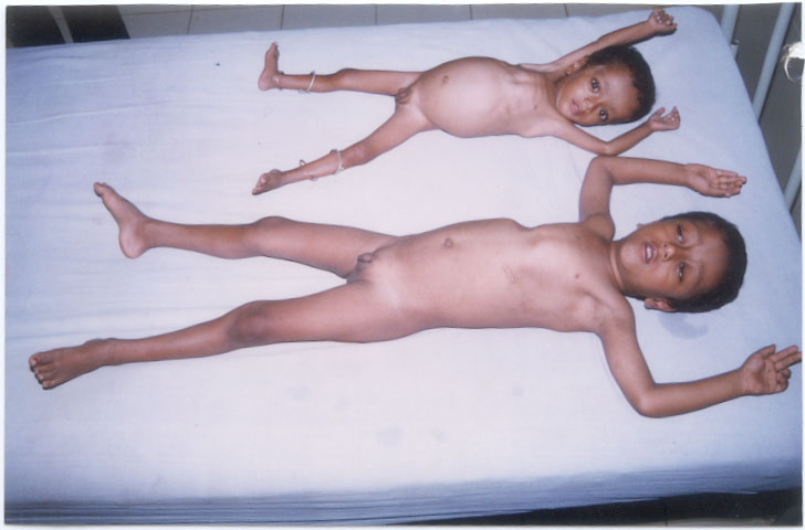
A 5-year-old boy, presented with history of inability
to walk and bear weight on lower limbs and difficulty in visualising
objects. This child was born to a consanguineously married couple. The
child had global developmental delay with an IQ of 50. The child was
short, with height of 92 cm and weight 12 kg, which were <5 centile on
NCHS charts. He had prominent eyes and gross deformity of the chest
(pigeon chest), frontal and parietal bossing, costochondral beading,
widening of the wrists and double malleoli (Fig. 1). On
examination, the liver was palpable 5 cm below the costal margin. Fundus
examination showed bilateral optic atrophy; hearing, which was assessed
by tympanometry and puretone audiometry was normal. Investigations
showed hemoglobin level of 11.4 g/dL, leukocyte count 9,600 cells/cumm,
platelet count 352,000/cumm, reti-culocyte count 0.2% and ESR 22 mm at
first hour. Serum calcium level was 7.5 mg/dL, phosphorus 2.5 mg/dL and
alkaline phos-phatase 259 U/L (normal 30-90 U/L). Other investigations
including renal parameters, electrolytes, blood gases and ultrasound
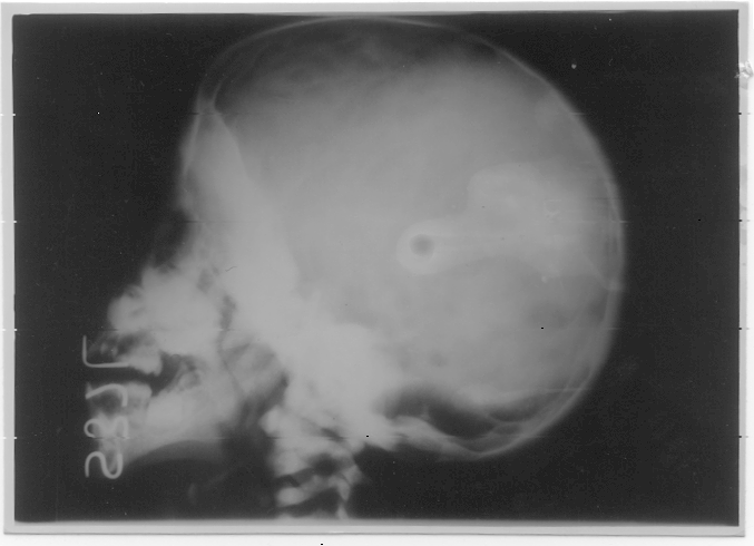
abdomen were within normal limits. Skeletal survey showed increased
density of all bones with obliteration of the medullary cavity in long
bones, which was diagnostic of osteopetrosis (Figs. 2 and 3).
The evidence of rickets in this case included clinical features,
radiological findings of fraying at the tip of ulna and biochemical
features.
 |
|
Fig. 1. Osteopetrorickets in two siblings
|
 |
|
Fig 2. Radiograph of skull (lateral) dense base
(case 1)
|
 |
Fig. 3. Chest radiograph showing dense sclerotic
rib cage (case 1) |
Case 2
The younger brother of our index case, aged 2 yr,
also had developmental delay with classical signs of rickets (frontal
bossing, parietal bossing, costochondral beading) and generalized
wasting with protruberant abdo-men and mild hepatosplenomegaly (Fig 1).
Optic fundus examination and hearing were normal. Biochemical features
included hypo-calcemia (7 mg/dL), hypophosphatemia (2.3 mg/dL) and
raised alkaline phosphatase (188 U/L). The hemoglobin level was 7.3 g/dL,
leukocyte count 9,300 cells/cu mm and ESR 40 mm at first hour. Renal
parameters and electrolytes were within normal limits. A diagnosis of
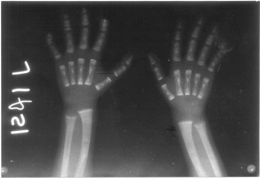
osteopetrosis with rickets was made based on radiological findings of
bone in bone appearance, fraying and cupping at the lower ends of the
bones of forearm (Fig. 4) and biochemical features.
 |
|
Fig 4. Radiograph of both wrist joints (AP view)
showing bone in bone appearance with evidence of rickets (case II)
|
Both cases were treated with high dose calcitriol and
oral calcium supplements(2) for 3 months with regular monitoring of
serum and urine calcium levels.
Discussion
Osteopetrosis (Albers Schonberg disease) is a rare
hereditary bone disease of heterogeneous pathophysiology occurring once
in 1-5 lakh children. The primary defect in all forms of the disease is
failure of the osteoclasts to reabsorb bone. A distinct form of
osteopetrosis in association with renal tubular acidosis and cerebral
calcification, due to carbonic anhydrase II deficiency, presents with
sensorineural hearing loss and psychomotor retardation.
Malignant infantile osteopetrosis is a rare autosomal
recessive disorder of osteoclast function characterized by abnormally
dense bone and failure of resorption of calcified cartilage. The major
clinical features derive from bony overgrowth of the marrow space and
compression of optic and auditory nerves, which pass through the major
foramina of the skull(3). Infants with this disease have failure to
thrive and are blind and deaf. They show hepatosplenomegaly, anemia and
thrombo-cytopenia due to bone marrow failure. Death usually occurs by
mid childhood as a result of bleeding, anemia or infection. Radiological
features are diagnostic with generalized osteosclerosis.
Rickets has been reported as a common and variable
feature of osteopetrosis(4). Its presence is enigmatic in light of the
markedly positive total body calcium balance associated with
osteopetrosis. The presence of rickets worsens the symptoms of
osteo-petrosis. Rickets is associated with increased lethargy,
irritability, poor feeding, growth retardation and pathological
fractures, and hence needs effective treatment.
In osteopetrosis the patients are often advised to
reduce calcium intake. In normal subjects a lower serum calcium
stimulates PTH secretion that acts on osteoclasts to cause bone
resorption and thus increase serum calcium. In osteopetrosis the
osteo-clasts do not respond to PTH and the responsibility for
maintaining calcium balance lies on the kidneys and intestine. Patients
on limited calcium intake and poor intestinal absorption due to
prednisolone, which is used for hematological complications, result in
reduced total body calcium and phosphate levels(5). Increased PTH
increases calcium reabsorption but at the expense of phosphate wasting.
Persistance of hypo-calcemia and hypophosphatemia results in inability
to mineralize newly formed chondroid and osteoid and the paradoxical
association of rickets and osteopetrosis.
Treatment options available include bone marrow
transplantation(6,7), glucocorticoid therapy (for hematological
abnormalities), and use of large doses of calcitriol, a potent bone
resorbing agent(2). Osteopetrorickets is a treatable condition and
should be treated by oral adminstration of calcitriol(8) and
liberalizing calcium intake especially if glucocorticoids are used for
control of hematological complications(2,8,9). We treated our patients
using high dose calcitriol (upto 8 µg/day) initially with a dose of 1
µg/day, gradually increasing to 2,4 and 8 µg/day every 15 days, over a
period of 3 months and oral calcium supplement of 320 mg/day.
The preferred treatment for infantile osteopetrosis
and its complications is HLA-identical bone marrow transplantation(6,7).
Nutritional support and calcium supple-mentation are necessary to treat
malnutrition and rickets.
Contributors: MLK diagnosed the case, drafted the
manuscript and guided the work and will act as its guarantor. PSM worked
up the case and reviewed the literature.
Funding: None.
Competing interests: None stated.
