|
|
Images in Clinical Practice Indian Pediatrics 2002; 39:593 |
|
Ash Leaf Macules |
|
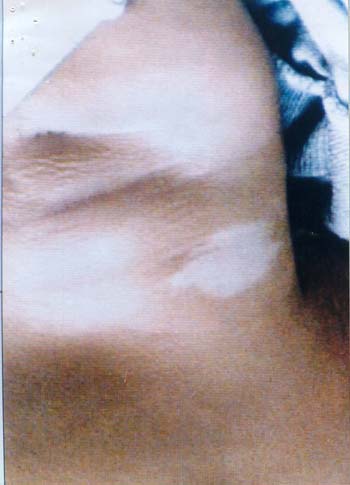
A 4-month-old product of non-consanguineous normal parents presented with three days history of recurrent seizures. Convulsions were of mixed type consisting of three to four episodes of generalized seizures interspersed with fifteen to twenty episodes of infantile spasms in a day. Examination revealed a normal weight baby with normal head circumference. Skin examination revealed three hypopigmented macules resembling ash leaves. Two were on low back and one was on anterior aspect of right shoulder (Fig. 1).
Fig. 1. Ash leaf macule on the anterior aspect of right shoulder. CT scan head revealed multiple subependymal tubers involving wall of frontal horn and body of lateral ventricle. There was evidence of calcification in the wall of lateral ventricle. EEG was suggestive of mixed seizure disorder. USG abdomen and Echocardiography were normal. Child was started on Sodium Valproate with significant control in seizures. Tuberous sclerosis is an autosomal dominant neurocutaneous syndrome with an incidence of 1:30,000. Bourneville and Brissaud first described it in 1880. Hypopigmented macules are seen in 50% of <2 year old. Rest of the clinical features like adenoma sebaceum, shagreen patches and sub ungual fibromas make their appearance latter in childhood. Prognosis for seizure control and mental retardation is guarded. More the number of tubers as was in this child, worse is the intellectual development. Ajay Kumar, Sarla Sharma. Department of Pediatrics, Hindu Rao Hospital, Delhi 110 054, India. E-mail: [email protected]
|
![]()