|
|
Case Reports Indian Pediatrics 2001; 38: 671-674 |
|||
|
Eosinophilia, Pleural Effusion and Cysticercosis - Unknown Association? |
|||
|
From the
Departments of Pediatrics and Radio- diagnosis*, Government Medical
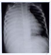
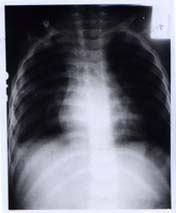
College, Chandigarh, India. Manuscript received: May 11, 2000; Initial review completed: July 4, 2000; Revision accepted: January 3, 2001. Pulmonary infiltration with eosinophilia (PIE) is a disease entity characterized by lung disease and eosionophilia. Common causes for it are tuberculosis, parasitic infestations, malignancy and connective tissue dis-orders(1). Many of the parasitic infections are mentioned as the cause for PIE. We report here a 2 year old child, who presented to us with PIE and subcutaneous cysticercosis. To the best of our knowledge, this association has not been reported in the literature. Case Report A 2 year-old-child, resident of Nepal was brought with history of fever and cough for 4 months and difficulty in respiration for two weeks. Two months back, he was started on antitubercular therapy for probable pneu-monia and positive family history of tuber-culosis, but there was no improvement and he was referred to our hospital. He belonged to a non-vegetarian family of low socioeconomic status and they often consumed pork. There was no hisotry of contact with animals. On examination, he was afebrile and vital signs were stable. There were three nodular swellings of size 1 ´ 1 cm on the trunk. Examination of the chest revealed findings suggestive of consolidation on the right side. Rest of the general physical and systemic examination was unremarkable. His hemo-globin was 10.2 g/dl, total leukocyte count 42,000/cu mm with absolute eosinophil count of 25681/cu mm. Chest radiograph and ultrasound revealed consolidation with pleural effusion on the right side (Fig. 1). Anti-tubercular drugs were continued and steroids were added (prednisolone 2 mg/kg). Pleural tap revealed 90% eosinophils, glucose 64 mg/dl and protein of 7.3 mg/dl. There were no malignant cells in pleural fluid and its culture was sterile. He was investigated for causes of pleural effusion with eosinophilia. Mantoux test was negative and gastric aspirate did not show acid fast bacilli. Toxocara canis antigen by ELISA, antifilarial antibodies and smear for microfilaria were negative. HIV serology, rheumatoid factor and antinuclear factor were also negative. Stool examination of the patient as well as his parents was normal. However, there was past history of passing worms in stools both by patient and his father. Ultrasound of subcutaneous swelling revealed presence of cysticercus, which was confirmed to be Cysticercus cellulosae on biopsy. Since tuberculosis seemed unlikely after all these investigations, his antitubercular drugs as well as steroids were stopped after 2 weeks. Radiograph of chest taken after one-week of therapy showed complete clearance (Fig. 2). He became afebrile and his cough also disappeared. He was given a 2 weeks course of albendazole. At 7 months follow up, there was no recur-rence of symptoms and absolute eosinophil count was only 1560/cu mm. The subcutaneous swellings over the trunk however remained same.
Discussion The entity "pulmonary infiltrates with eosinophilia" was coined by Reeder and Goodrich for all the disorders having pulmonary infiltrates with eosinophilia like Loeffler’s syndrome, prolonged pulmonary eosinophilia, pulmonary eosinophilia with asthma, tropical eosinophilia and panarteritis nodosa. Later on, it was appreciated that pulmonary infiltrates with eosinophilia can occur in absence of peripheral blood eosinophilia. As a result, the term eosinophilic pneumonia was used to include all disorders characterized by infiltration of lungs with eosinophils, with or without an excess of eosinophils in the peripheral blood(1). Our child had involvement of lungs with peri-pheral as well as lung eosinophilia. Pathophysiologically, this pneumonia consists of filling of alveoli with eosinophils, large mononuclear cells and by an interstitial infiltrate of eosinophils, lymphocytes and plasma cells. Accumulation of eosinophils in the lower respiratory tract is thought to be due to release of chemotactic factors by the lung(2). Eosinophils are known to cause destruction of tissue. Corticosteroids have been shown to have a dramatic effect on the function of eosinophils in vitro(3). Our patient also had dramatic improvement with steroids. Various parasites known to cause eosinophilic pneumonia are toxocariasis, filiariasis, ascariasis, paragonomaniasis and hydatidosis. In a child who has pleural effusion and presence of eosinophilia in the peripheral blood, tuberculous or malignant pleural effusion is less likely and the probability of it being benign is higher(4). Our patient probably had Loefler’s syndrome, i.e., mild disease with transient chest involve-ment, as chronic eosinophilic pneumonia requires repeated or prolonged use of corticosteroids. Temporal relationship with presence of subcutaneous cysticercosis is interesting. Intestinal parasitism is less commonly associated with eosinophilia as compared to tissue invading parasites(2). Cysticercosis, a tissue invading condition has not been reported as a cause for pleural effusion in the existing literature, though it is known to cause eosinophilia in its early stage(2). Presence of peripheral as well as pleural fluid eosinophilia with pneumonia and temporal presence of subcutaneous cysti-cercus cellulose and complete clearing of the lung picture after additon of steroids points towards cysticercus cellulosae being the cause of these clinical features by hypersensitivity reaction. Although, hypersensitivity pneu-monia usually presents as diffuse, poorly defined nodular radiodensities, even pleural effusion, atelectasis, calcification and local-ized radiodensities have been mentioned as rare manifestations of this pneumonia(5). Infection with Taenia solium is frequently found in Eastern Europe, China, Pakistan, India, Mexico and central and South America. Usually, human being is the definitive host for Taenia solium and the parasite stays in the small intestine attached to mucosa, causing abdominal symptoms and weakness. In cysticercosis, humans are the intermediate host by ingestion of eggs in contaminated food or water, oral transmission through unclean hands and internal autoinfection by regurgitation of eggs into the stomach(2). Cysticercus cysts may develop anywhere in the body, but show predilection for brain, meninges, eyes, muscle, heart, liver, lungs, oral cavity and skin. Subcutaneous nodules are usually asymptomatic, round, rubbery and usually of size 1-2 cm. Allergic phenomenon leads to erythema, swelling, blood eosino-philia and raised serum IgE levels(6). The same allergic phenomenon may have been responsible for eosinophilic pneumonia in our patient, which got cleared rapidly with the addition of steroids. Treatment of subcuta-neous cysticercosis is surgical, though people have used praziquental and albendazole(7). Keeping the possibility of cysticercosis as cause for pleural effusion with eosinophilia in endemic areas will spare the patient from unnecessary investigations. Contributors: MS did data collection, writing and drafting of the paper and will act as guarantor for this paper. SM helped in patient management and data collection. VP helped in drafting the paper. SK diagnosed the condition by ultrasound of the patient. Funding:
None.
|
![]()