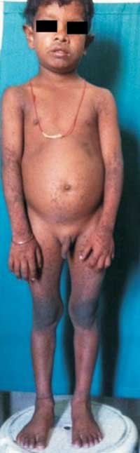
A 6-years-old boy presented with multiple
dark, raised lesions all over the body since 1 year (Fig.
1a). The disease began with discrete
hyperpigmented hyperkeratotic papules over the knee and
elbows which later progressed to involve the preauricular
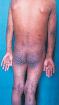
region, ear lobe, neck, both flexor and extensor aspect of
upper limbs and lower limbs and buttocks (Fig. 1b).
There was history of photo-exacerbation of the lesions. The
child complained of pruritus and difficulty in sitting due
to pain because of multiple hyperkeratotic lesions over the
buttocks. There was presence of palmar pits. Oral mucosa and
nails were normal. His 4-year-old sibling also presented
with similar lesions over the knees and elbows. A clinical
diagnosis of Darier-White disease was made.
Histopathological examination from punch biopsy of a lesion
showed acantholysis along with classical dyskeratosis and
hyperkeratosis in the epidermis.
 |
 |
| (a) |
(b) |
|
Fig. 1 Multiple
hyperkeratotic lesions including (a) knees and
elbows, and (b) buttocks.
|
Darier-White disease or keratosis
follicularis is said to occur as a result of mutation in the
ATP2A2 gene located on chromosome 12q23-24.1,
responsible for coding sarco/endoplasmic reticulum calcium
ATPase type 2 (SERCA2). It is characterized clinically by
hyperkeratotic papules distributed mostly on the seborrheic
areas of the body. Nail involvement is characterized by
V-shaped nicking at the distal aspect of the nail bed,
longitudinal red and white alternating bands, and subungual
hyperkeratosis. Mucosal membrane involvement may occur as
white papules on the buccal mucosae, palate, and gingiva
with a cobblestone appearance. Palmoplantar involvement
usually presents as discrete, punctate keratoses that appear
as small, hyperkeratotic papules or small, centrally
depressed pits. In flexures like axilla and groin, the
lesions may become large exuberant growth. They may get
infected due to constant maceration resulting in malodorous
purulent discharge. Heat, sweat, humidity, sunlight, oral
corticosteroids and mechanical trauma have been reported to
exacerbate this condition.
Conventional therapy for severe disease
still relies greatly on oral retinoids. Acitretin is
effective at 0.6 mg/kg/day, the hyperkeratosis is reduced
and papules are flattened. Basic measures include use of
sunscreens, cool cotton clothing, and avoidance of hot
environment. Moisturizers with urea or lactic acid can
reduce scaling and hyperkeratosis. Surgical treatment
includes dermabrasion, carbon dioxide laser, and the erbium
YAG laser. The condition runs a chronic relapsing course,
with exacerbations throughout life.


