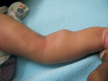
A 21-day-old male infant presented with swelling over left upper limb for
3 days. Examination revealed a firm swelling of 3×4 cms over the proximal
ventral aspect of left forearm and linear swellings over the medial aspect
of left upper arm (Fig. 1(a)). Systemic examination
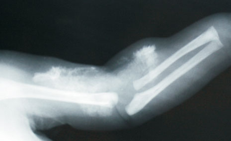
and routine blood workup was normal. Ultrasound and X-ray of the
swelling revealed subcutaneous calcification (Fig. 1(b)).
This baby had asphyxia at birth, developed seizures and hypocalcemia on
2nd day of age, hence was treated with 10% calcium gluconate intravenously
for 4 days and discharged with oral calcium supplementation. A diagnosis
of iatrogenic calcinosis cutis was made.
 |
 |
|
(a) |
(b) |
|
Fig. 1 (a) Showing swelling in the upper
part of the left forearm, (b) X-ray limb showing subcutaneous
calcification. |
Calcinosis cutis is characterized by abnormal deposits
of calcium salts in the dermis and/or hypodermis due to transient
elevation of the local calcium concentration after intravenous
administration and local trauma. The differential diagnosis includes
cellulitis, osteomyelitis, arthritis, abscess, periostitis,
thrombophlebitis and myositis ossificans. Calcinosis cutis is
differentiated from cellulitis and abscess by absence of signs of
inflammation and characteristic roentgenographic findings. Myositis
ossificans usually appears within the first decade of life as spontaneous
or injury-induced exacerbations. The lesions are characterized by painful
swellings in soft connective tissue, including tendons, ligaments, fascia,
and skeletal muscle. No specific treatment is required for iatrogenic
calcinosis cutis as it resolves spontaneously within three months.


