|
|
Images in Clinical Practice Indian Pediatrics 2000;37: 797-798 |
||||
|
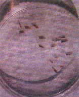
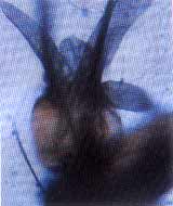
Urogenital Myiasis |
||||
|
Myiasis in an extremely rare condition produced by larval stages of flies. The condition resulting from larvae requiring living tissues of animal or human beings to develop is known as obligatory myiasis and that in the wound as semispecific myiasis. Myiasis can be cutaneous, ocular, intestinal and urogenital depending on the organ involved. The larvae of the flies are attracted to the draining infections or to the clothing soiled with urine or faeces. They crawl into the natural orifices when an infected person sleeps on the ground or is any way exposed to the flies. Individuals living in poor sanitary conditions are particularly susceptible. The exact source of urogenital myiasis is not clear. Possibly, the larvae gain access into the urinary bladder through urethral orifice, lodge and grow there and subsequently get excreted in the urine.
B.K. Rath, P.K. Das,
|
![]()