|
|
Case Reports Indian Pediatrics 2000;37: 786-789 |
|||
|
A Child with Prolonged Pyrexia and Peripheral Desquamation: Is it Kawasaki Disease? |
|||
|
Sudeshna Mitra
Kawasaki disease (KD), an acute systemic vasculitis of young children is diagnosed if five criteria out of six laid down by the Japan Kawasaki Disease Research Committee are present(1). If coronary abnormalities can be demonstrated, then presence of any four criteria suffices for diagnosis. However, several children have been reported in the literature with coronary abnormalities pathognomonic of KD yet not fulfilling the minimum criteria(2). They have been clubbed under the diagnosis of atypical KD (AKD). Early recognition of these cases is important since they may suffer myocardial infarction or an "unexplained" death years later(3). Some cases have actually been diagnosed at autopsy(4). We report here a case of AKD with unusual presentation.
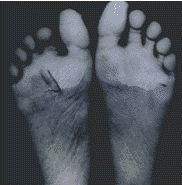
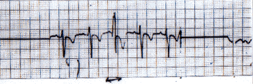
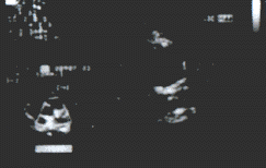
A 10-year-old boy was admitted with high fever and sore throat for 4 days, breathlessness for 1 day and palpitations for a few hours prior to admission. On examination he was febrile, had tachycardia, tachypnea and a congested throat. He was diagnosed to have a right sided consolidation. Chest X-ray revealed bilateral small pleural effusions in addition. He had polymorphonuclear leukocytosis while the hemoglobin and platelet counts were normal. Electrocardiography showed sinus tachycardia. C-reactive protein was positive, throat swab and anti-streptolysin O titer were non-contributory. He was started on parenteral amoxycillin clavulanate. A pleural tap was done which yielded pus. There were Gram positive cocci on smear; culture was sterile. He was put on a thoracostomy drain and given 2 weeks of ceftriaxone and vancomycin since he was allergic to penicillin. The fever, however, did not subside. On the 10th day of hospital stay, ultrasound and CT chest were done. These showed thick pleural septations, so he under-went thoracotomy decortication. Fibrinous exudates with adhesions seen per-operatively were cleared. Post-operatively, fever, and tachycardia persisted. Pleural specimen sent for culture was sterile and was consistent with fibrinous pleuritis. Investigations repeated while the child was off antibiotics failed to localize the cause of fever. Chest X-ray showed complete resolution of consolidation and effusion. There were no vegetations on echocardiography and the coronaries were normal. On the 16th day of admission des-quamation was noticed on the fingertips. Platelets counts repeated at this time with a suspicion of AKD, rose from 3 lakh/mm3 in the third week of illness to 12.5 lakh/min3 in the fourth week. Another week down the course he had peeling of skin of the soles (Fig. 1 ) which progressed proximally. A few days later irregularity of pulse was noted and ECG showed occasional ectopics (Fig. 2 ). In view of unexplained fever, acral desquamation, arrhythmia and thrombocytosis, AKD was diagnosed. He was given IVIG 400 mg/kg/d for 4 days and started on high dose aspirin. Fever subsided although tachycardia and ectopics continued. He was discharged on low dose aspirin. On follow-up 2 months later echo-cardiograph showed dilatation (5.6 mm) of the origin of the left main coronary artery (Fig. 3 ). Holter revealed an average heart rate of 110/min and multiple episodes of sinus tachycardia with rates of 120-167/min. Approximately 4% of the beats were ventricular ectopics-there were no runs. The irregularity of pulse was present till 7 months later after which it decreased markedly. At 12 months follow-up there was only an occasional missed beat. Repeat echo done at this time showed resolution of coronary abnormality. However, thallium scan 18 months after the illness suggested ischemia of the anterior wall.
Fig. 1. Peeling of the soles
Fig. 2. ECG showing ventricular ectopic
Fig. 3. Echocardiogram showing dilatation of left main coronary artery (arrow).
This child had a very atypical course. Less than 20% of cases of KD occur in children above 5 years age. Onset after 8 years is said to be rare(5). Even AKD has been seen mostly in infants(6). Rarely systemic or focal infection with group A streptococcus or Staphylococcus aureus can present with some features of KD(6). Although, blood culture was negative in our case there was clear evidence of empyema chest. Initial presentation with infection at some site, pleural effusion and pulmonary infiltrates have all been described in the context of AKD(7). However, the course was rather prolonged in this child before the features of KD manifested. Superantigens (streptococcal and staphylococcal toxins) implicated in the pathogenesis of KD(8) might have been responsible. Periungual desquamation or thrombocytosis if present with prolonged unexplained fever mandates echocardiography(9). If ignored in view of their non-specificity, the outcome may be fatal(3). Several ECG changes have been observed in KD. Although, our patient had only occasional ventricular ectopics, this arrhythmia persisted for long. Usually, such changes normalize by 6 months(10). Aneurysms are said to appear between 9-15 days of illness but in the case described coronary artery dilatation was detected 2 months after illness which is uncommon(6). This occurred inspite of IVIG administration hence reinforcing the urgency of starting IVIG early. To be of benefit, IVIG should ideally be given within 10 days of onset of illness(5). The effectiveness after this period is not clearly known, but the anti-inflammatory effect may do some good. Also, it has been seen that if IVIG treated patients develop coronary abnormality they were more likely to have resolution within 2 years than those treated with aspirin alone(11). Myocardial infarction may occur till many years later but usually occurs within the first year(12). Our patient continues to have minor irregularity of pulse and there is a possibility of his having sustained an asymptomatic myocardial infarction as has been described earlier(12). Repeated attempts have been made over the years to evolve a scoring system to determine which cases are likely to develop coronary artery abnormalities(13). Amongst the different parameters, persistence of fever beyond 14 days has been reiterated. Our patient had prolonged fever, signs of myocarditis and thrombocytosis all of which were high risk factors for developing coronary artery abnormalities. Although young age has been mentioned as a risk factor, a Japanese study reported high prevalence of cardiac sequelae both in infants and children over 5 years(14). In another study 18.5% patients with coronary artery abnor-mality failed to meet the diagnostic criteria of KD(9). Ours is probably one such case. In a disease where diagnosis is based on fulfilment of certain clinical criteria, it is difficult to label a case ‘atypical’ where criteria are not met. A somewhat similar case has been recently reported where a suspicion of KD arose when a 2 year-old boy with chickenpox had peeling of fingers(15). In the case discussed here, although the diagnosis was in doubt when IVIG was administered, echocardiography and thallium scan subsequently have proved the diagnosis since coronary artery abnormalities and myocardial ischemia are extremely odd in other pediatric conditions. This report should serve to encourage physicians to consider a diagnosis of AKD in a febrile child in whom some but not all of the features of KD are present and persistence of fever is unexplained. Establishing a diagnosis of KD, especially AKD, can be a major clinical dilemma, because signs like prolonged fever, rash, conjunctivitis, pharyngeal congestion are very non-specific. Added to this is the lack of a confirmatory laboratory test. In an odd case, like the one described, it may be difficult to decide in favour of IVIG therapy considering the expense. At the same time there is some urgency in starting treatment early.
|
![]()