|
|
Correspondence Indian Pediatrics 2007; 44:53-54 |
|||
|
Primary Tuberculosis of Mandible |
|||
|
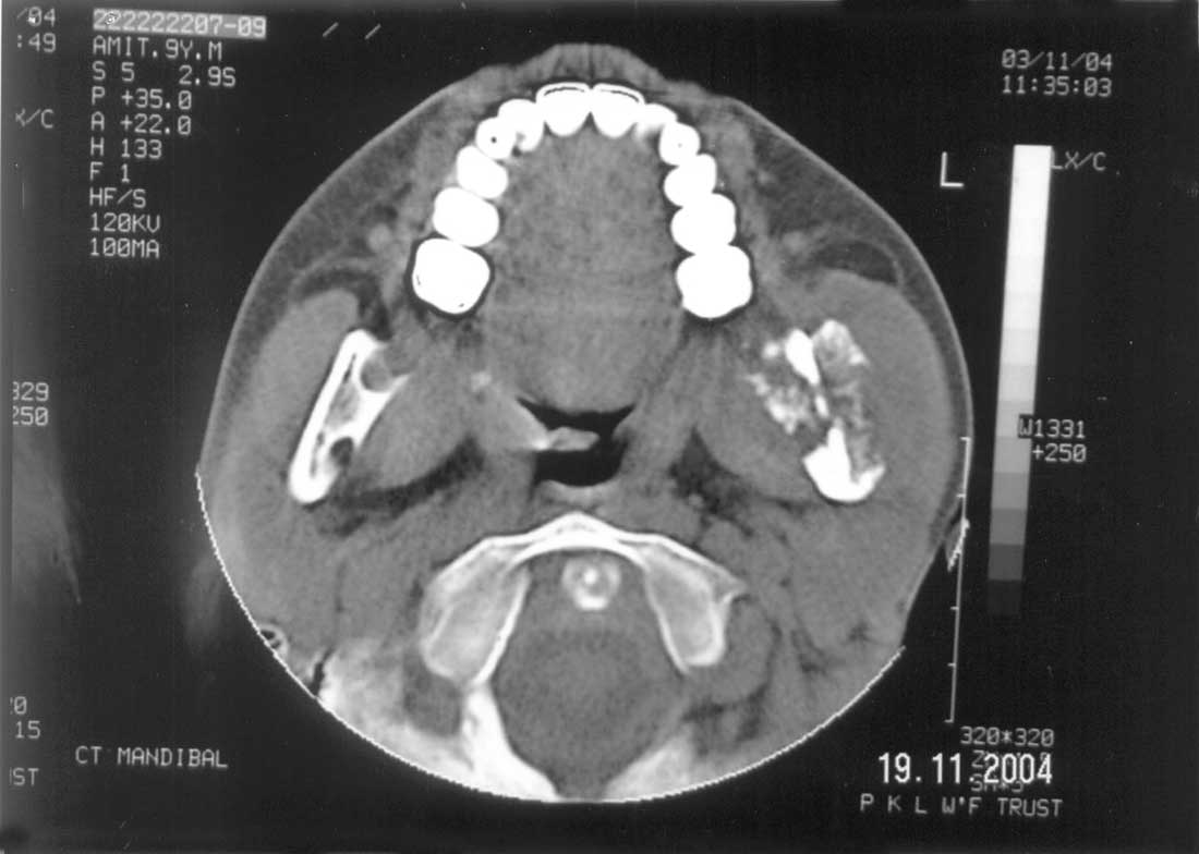
Routine laboratory tests were grossly unremarkable. Radiograph of mandible revealed expansile osteolytic lesion involving angle of left mandible while the chest X-ray done to look for any evidence of primary focus, was normal. Gastric aspirates for acid-fast bacilli (AFB) and tuberculin skin test were negative. Non-contrast CT scan of mandible (Fig.1) revealed destruction of inner as well as outer wall of the angle of left mandible due to expansile osteolytic mass lesion. Osteoblastic activity was also seen as new bone formation around the lytic lesion. Breach in periosteum with extension of expansile lesion into surrounding soft tissue was also noted. CT findings were reported as malignant bone tumor. Fine needle aspiration smear from the swelling showed necrotizing granulomatous inflammation consistent with tuberculosis but Ziehl Neelsen staining for AFB was negative. Standard antitubercular therapy for bone tuberculosis was started. At 2 weeks follow-up some reduction in size of swelling was noticed and swelling completely disappeared at 2 months follow-up. Bone tuberculosis is a relatively uncommon form of extrapulmonary tuberculosis seen in approximately 1% of children with tuberculosis(1). It more frequently seen in children as compared to adults because epiphyseal region of the bones is highly vascularized in infants and young children. Most reported cases of mandibular tuberculosis were secondary to tuberculous focus elsewhere in the body. To best of our knowledge, only 4 cases of primary mandibular tuberculosis have been reported(2-4). Three routes of infection to the mandibular bone are postulated. One is direct transfer of infected material through a carious tooth, a post extraction socket or mucosal wound. Other two are direct extension from local soft tissue lesion to the underlying bone and hematogenous route. In our patient, a slight wound in the oral cavity or gingivitis might be the entry site, though there was no history of extraction of carious tooth or trauma. A similar route of entry had been postulated in another reported case(4). Mukesh Kumar Gupta, Acknowledgement We thank Dr. Devidayal for his assistance in clinical assessment and patient management.
|
![]()