|
|
Letters to the Editor Indian Pediatrics 2005; 42:86-87 |
||
|
Combined Esophageal Duplication Cyst with Bronchogenic Cyst |
||
|
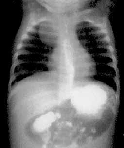
Ultrasonogram revealed cystic lesion in the right upper chest with internal echoes. Barium swallow showed a mass lesion causing smooth indentation of the esophageal outline (Fig. 1). At surgery, a cyst was identified in the posterior mediastinum, which was para-esophageal in location extending into the superior mediastinum. After excision, clinical improvement was rapid and child is well 8 months later.
The 4 × 5 cm ovoid mass showed mucoid material on cut section. A unilocular cyst was seen with smooth inner wall. Microscopic sections showed esophageal duplication cyst lined by non-keratinizing squamous to cuboidal epithelium with goblet cells. The muscular wall showed distinct inner and outer muscularis propria. No ganglion cells were seen. The adjoining fibroconnective tissue showed a very tiny bronchogenic cyst lined by pseudostratified ciliated columnar epithelium to an attenuated epithelium. Wall showed only fibroconnective tissue with subjacent mucus glands and islands of cartilage. The bronchogenic cyst was only evident microscopically. There was no communication between the two cysts. The term Bronchopulmonary Foregut Malformation (BF) was coined by Gerle et al in 1968 to describe pulmonary sequestrations having patent congenital communications with the upper GI Tract(1). The primitive foregut gives rise to the pharynx and lower respiratory tract as well as upper GI tract. The most common foregut cysts, the bronchogenic and the esophageal duplication cyst represent abnormal budding of the vertebral and dorsal primitive foregut respectively(2), indicating their common origin from the primitive foregut. These foregut cysts are lined by ciliated epithelium which occurs both in early tracheo-bronchial tree and oesophagus. Bronchogenic cysts and extralobar sequestrations may originate from the embryonal foregut and thus may have a close embryologic relationship(3). Congenital Cystic Adenomatoid mal-formation (CCA), Congenital Lobar Emphy-sema (CLE), pulmonary sequestrations and bronchogenic cysts are anomalies of foregut duplication which can present as cystic masses in the chest. They usually present as separate lesions but very rarely can occur in combination(4). The clinical presentation is often due to compression symptoms or respiratory infection. Imaging with chest X-ray, ultra-sound, barium swallow and now CT or MRI delineates the dimensions and relations. Surgical resection is curative for this condition. Smrita Dorairajan,
| ||
|
References | ||
|
|
![]()