 |
Images in clinical Practice Indian Pediatrics 2003;40: 62 |
|
Chronic Bullous Dermatosis of Childhood |
A seven-year-old boy presented with multiple vesicobullous lesions over the lower abdomen, genitalia and both extremities for four years. The lesions were discrete, tense, and the ‘bulla spread sign’ was negative. They were lying over a non-erythematous base while the surrounding skin was variably pigmented, denoting areas of prior healed lesions (Fig.1). Excoriation marks revealed the itchy nature of the disease. Mucous membranes, hair and nails were uninvolved. The course of the disease had been waxing and waning. New lesions always appeared in vicinity of the older healed ones. Intermittent courses of oral steroids had helped in temporary partial remission, only to resurface again. The patient was started on dapsone 25 mg per day and a dramatic, though partial response was evident by the end of first week. At the end of second week, the dose was escalated to 50 mg per day, and the patient has been in complete remission since.
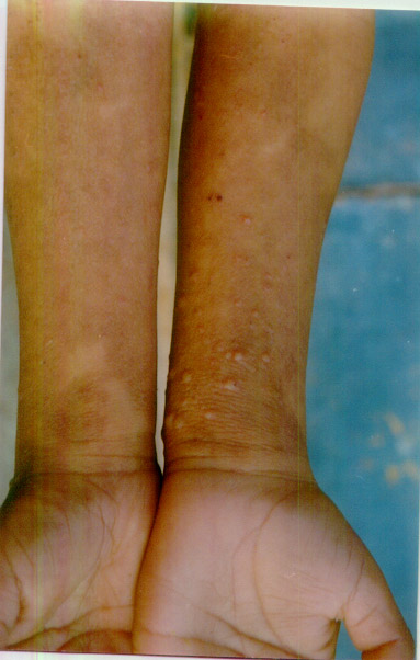 |
| Fig.1 Multiple grouped vesicobullous lesions seen on the flexor aspect of forecarms with surrounding post-healing pigmentation. |
Chronic bullous dermatosis of childhood is considered to be an autoimmune, acquired, self-remitting vesicobullous disease with a mean duration of 4-4.5 years. It characteristically involves lower abdomen, genitalia, lower extremities and periorificial areas, with a tendency for newer lesions to appear in proximity to the prior lesions, sometimes forming the characteristic ‘cluster of jewels’. Sulfones form the mainstay of therapy, while additional corticosteroids may be required in some cases.
Ram Gulati,
Gautam Bagga,
Department of Skin, STD & Leprosy,
Mahatma Gandhi National Institute of Medical Sciences,
Sitapura Institutional Area, Jaipur, Rajasthan.
E-mail: dr_ramgulati@yahoo.com
![]()