|
|
Images in Clinical Practice Indian Pediatrics 2002; 39: 97 |
|
Brush Field Spots |
|
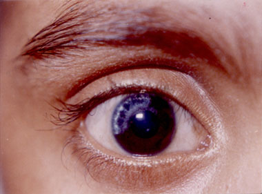
An 11-year-old boy with dysmorphic facies was referred to Ophthalmology Outpatient Department for evaluation of whitish spots in both eyes present since birth. The child was a product of non-consanguineous marriage, 4th in birth order with maternal age of 30 years at time of conception. The child had a global delay in the developmental milestones. The physical appearance was suggestive of Down’s syn-drome with brachycephaly, flat occiput, flat face, low set ears, high arched palate, flat nasal bridge and hypotonia. The chromosomal analysis confirmed trisomy 21. The ophthal-mological examination revealed telecanthus and mild blephritis. Cornea was normal in both eyes; iris of the right eye had multiple white spots in the supero-temporal quadrant in the periphery and along the pupillary margin with speckling suggestive of Brushfield spots (Fig.1). The left eye showed superior sectoral hypotrophy with which there was an accent of Brushfield spots. Vision could not be assessed, as the child was not cooperative. Both eyes had anterior subcapsular cataract. Patient was myopic and had no fundal abnormality. Brushfield spots are white or yellow colored spots seen on anterior surface of the irides. They are present in 85% of blue or hazel eyed patients with trisomy 21. They can be arranged concentric to the pupils, mid periphery or along the collarette. Only 17% of the brown iris with Down’s Syndrome have Brushfield spots as they are obscured by the anterior concentration of the pigment cells. Indian’s have dark pigmented eyes making Brushfield spots less appreciable in them. The Brushfield spots need to be differentiated from stromal condensations named "Kunkmann Wolffian" bodies, which are seen in 15% of normal, light colored iris. These are less distinct, less numerous and more peripheral than Brushfield spots with no associated facial dysmorphism.
Fig. 1. Brushfield spots in the right eye seen in the peripheral iris and along the pupillary margin. Usha Langan*, |
![]()