|
Images in Clinical Practice |
Indian Pediatrics 2000;37: 102 |
Subcutaneous Fat Necrosis of Newborn |
A one hour old boy presented with blue discoloration of body. He was a full term vagi-nal delivery conducted in a private nursing home. The baby did not cry immediately after birth and was resuscitated. There was history of obstructed labor and pre-eclampsia in the mother. The investigations revealed no evidence of sepsis. The child was given appropriate treat-ment in the form of oxygen, intravenous fluids and antibiotics. There was clinical improvement in the condition of the child by day 3.
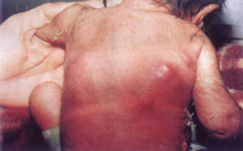
Fig. 1. Photograph showing multiple, reddish, nodular, swellings over the back in the right infrascapular area On 10th day of life, the child developed multiple, reddish, nodular, non tender, fluctuant swellings over the back in the right infrascapular area and induration in the left infrascapular area (Fig.). Excisional biopsy from one of the swellings showed unhealthy granulation tissue along with fat deposits in crystalline form with both acute and chronic inflammatory pathology suggesting subcutaneous fat necrosis of new-born. The liquefied material collected during the biopsy showed no growth of pyogenic organ-isms on culture. Within one month, all these swellings disappeared without leaving any scar. However, there was appearance of another simi-lar swelling in the right gluteal area, that also disappeared within 15 days. On follow up for another one month, the baby was found to be normal. Subcutaneous fat necrosis of newborn is a benign, self-limiting condition and carries a good prognosis. It is usually associated with hypercalcemia; which however was not there in the present case. Glucocorticoids and with-drawal of Calcium and Vitamin D are the treat-ment of choice for hypercalcemia. K.K. Locham and G.S. Parmar, |