An II-year-old boy presented with a sudden onset of left sided
hemiparesis. He had a similar episode 5 years back preceded by left
sided focal convulsions. The hemiparesis recovered within a few
months. He was studying in 6th standard and his school performance was
satisfactory.
 |
|
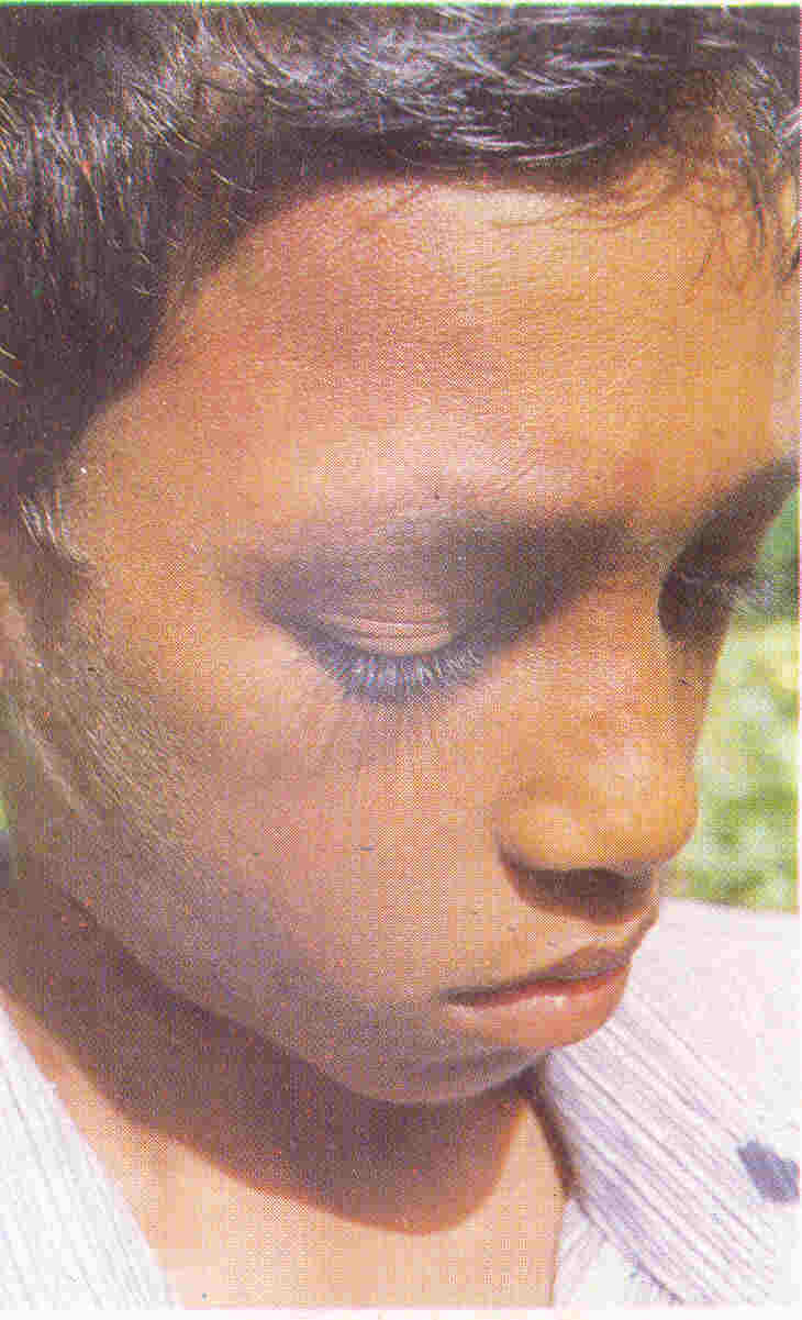
Fig. I. Close up of the face showing a capillary hemangioma on the right
forehead extending from below the hairline, involving the upper part of
right eyebrow and going up to the right side of the nasolabial fold. |
His growth and development was also normal. On examination he had a
capillary hemangioma involving the right side of forehead and upper
eyelid, measuring 8 cm x 5 cm (Fig. 1) and left sided upper
motor neuron palsy involving the face, upper limb and lower limb. Rest
of the examination was within normal limits. X-ray skull revealed
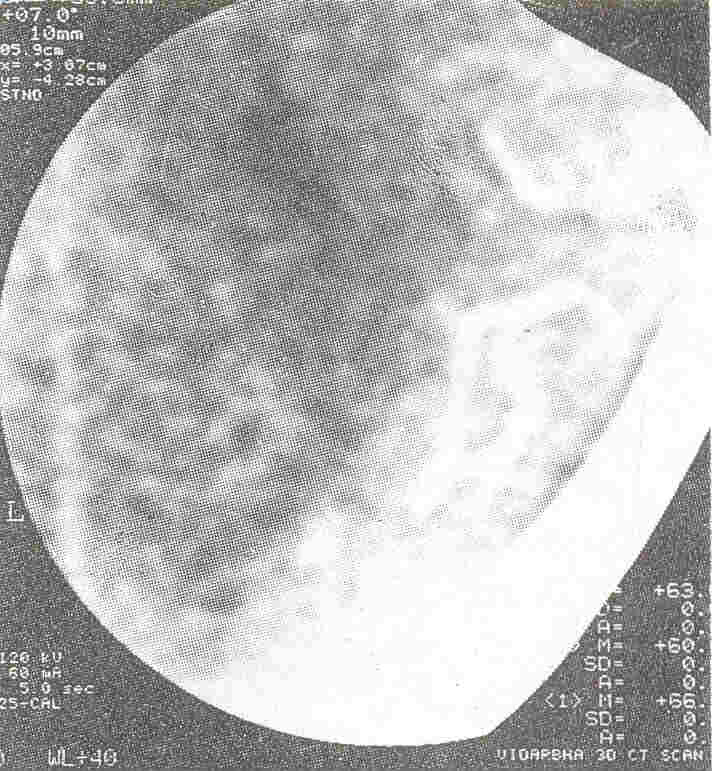
doubtful rail track calcification in the occipital area. CT Scan
revealed unilateral right parieto-occipital tram like and serpentine
calcification following convolutions suggestive of a calcified
cerebral hemangioma (Fig. 2).
 |
|
Fig. 2. Magnified view of the CT scan showing unilateral right parietocciptal
tram like and serpentine calcification following
convolutions in the supratentorial compartment. |
Pushpa Chaturvedi, Pradeep Sahare,
Nandkumar Banait,
Kasturba Hospital,
MG1MS Sevagram,
Wardha 442102,
Maharashtra, India.
