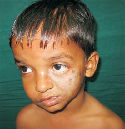
A six-year-old boy presented with skin lesions, initially started on the
left cheek at 2 years of age, then spread to left lower eyelid and
forehead. On examination, diffuse sclerosis on the left cheek extending
to left lower eyelid was noted. Hypoplasia of left half of the face and
deviation of mouth and lips to left side were noted, with loss of
eyelashes in the left lower eyelid (Fig. 1 and 2).
Investigations show normal blood counts, renal function tests, and liver
function tests. Rheumatoid factor and antinuclear antibody were
negative. Skin biopsy shows features suggestive of early morphea. CT
scan of brain was normal. CT scan of face shows hypoplastic left
maxillary sinus, left hemi-mandible, atrophy of the left hemi-facial
muscles and subcutaneous fat.
 |
|
Fig.1 Marked hypoplasia of the left half of the face with
deviation of lips toward left side and loss of eyelashes in left
lower eyelid.
|
Parry-Romberg syndrome is an uncommon degenerative
condition characterized by a slow and progressive atrophy of facial
tissues. A sharp demarcated line between normal and abnormal skin called
coup de saber develops. The diagnosis is clinical and based on
characteristic cutaneous and soft tissue findings.
 |
|
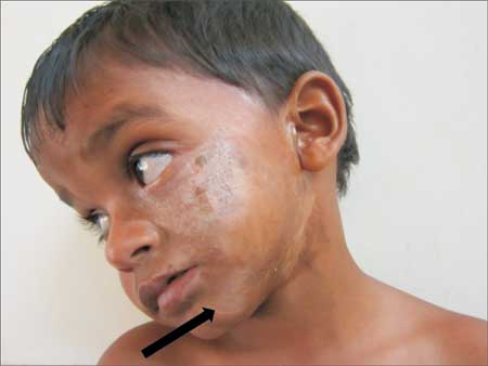
Fig. 2 A big linear scar (coup de
sabre) in the left side of mentum region.
|
Differential diagnoses include hemifacial microsomia
(first and second branchial arch syndrome) and its variants, such as Goldenhar
syndrome, post-traumatic atrophy and partial lipodystrophy (Barraquer-Simon
syndrome). Hemifacial microsomia and Goldenhar syndrome are congenital
and non-progressive. In post-traumatic atrophy, history of trauma will
be present. Lipodystrophy is usually bilateral and involves primarily
the adipose tissue. Parry-Romberg Syndrome and localized scleroderma may
represent differential spectra of same disease. Parry-Romberg syndrome
will have hemifacial atrophy of the skin and tissue below the forehead,
where as localized scleroderma is generally located in the fronto-parietal
scalp and/or the paramedian forehead; it may extend down the face as
well.
It is an auto-limitable condition and there is no
cure. The treatment is usually based on reposition of adipose tissue.


