|
|
Case Reports Indian Pediatrics 2007;44:-931-933 |
||||
|
Kniest Syndrome |
||||
|
Subramanian Subramanian From the Department of Radiodiagnosis and Department of Ophthalmology*, All India Institute of Medical Sciences, Ansari Nagar , New Delhi 110 029, India. Correspondence to: Dr Subramanian Subramanian,
Department of Radiodiagnosis, All India Institute of Medical Sciences,
Ansari Nagar, New Delhi 110 029, India. Manuscript received: January 19, 2007; Initial
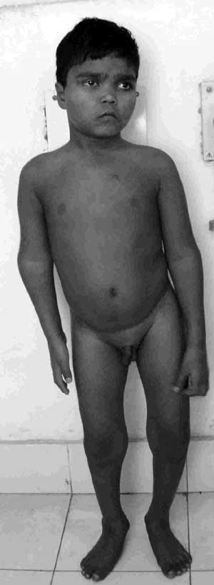
review completed: February 28, 2007; Abstract: A 12-year-old male child presented with bilateral visual loss and short stature. He had dysmorphic faces, barrel shaped chest and short limbs with enlarged peripheral joints. In addition he had bilateral retinal detachment with secondary cataract formation. Skeletal survey revealed irregular platyspondyly, hypoplastic femoral head and enlarged epiphysis of long bones with cloud like calcification. Radiological features were diagnostic of Kniest syndrome. The child underwent pars plana lensectomy and a vitreo-retinal surgery with silicon oil infusion in the right eye for retinal detachment. Key words: Kniest syndrome, Retinal detachment .Kniest syndrome was first described by Wilhelm Kniest in the year 1952(1). Children affected by this syndrome have round faces, barrel shaped kyphotic trunk, disproportionate dwarfism, and enlarged joints with restricted movements(2). They may present with visual loss due to retinal detachment and myopia or with recurrent otitis media(2). The disease is inherited in autosomal dominant manner. We present a rare case of Kniest syndrome with ocular involvement in this report. Case Report A 12-year-old male child presented with loss of vision in both eyes and short stature. He had visual loss in left eye for four years and in the right eye for one year. He was born to non-consanguineous parents and his siblings were normal. He had normal milestones and intelligence. On examination, the child had a flat round face with epicanthal folds, depressed nasal bridge, and high arched palate. The chest was barrel shaped and there was kyphoscoliosis in lower back (Fig. 1). The limbs were shortened and knee, elbow, wrist and ankle joints were enlarged with limited mobility. His height was only 104 cm. Based on the clinical features Moriquio’s disease was suspected.
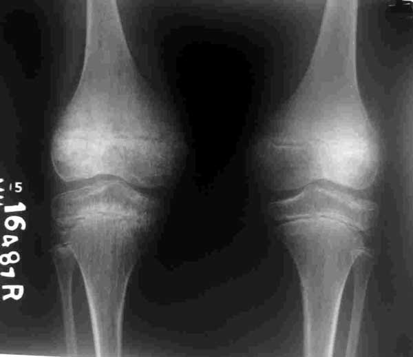
Ocular examination revealed bilateral cataract. The cornea was clear on slit lamp examination. The intraocular tension was normal. As the retina could not be evaluated due to the presence of cataract, sonogram was done. Ocular sonogram revealed funnel shaped retinal detachment in right eye and closed funnel retinal detachment in left eye. Skeletal survey revealed irregular platyspondyly with scoliosis in the lumbar vertebra. The pubic bones were hypoplastic and pelvic inlet was smaller in size. The ossification centre for femoral head was smaller in size and fragmented. The epiphysis of the lower end of femur was enlarged and metaphysis was flared. Cloud like calcification was seen around the growth plate (Fig. 2). The epiphysis in elbow, wrist and ankle joints also showed similar change. Radiograph of hand revealed flattened epiphysis of metacarpal bones with enlargement of both ends of metacarpal and phalanges. Joint space was reduced in the small joints of hand. The skull radiograph was normal. The radiological features are diagnostic of Kniest syndrome. The child underwent pars plana lensectomy and a vitreo-retinal surgery with silicon oil infusion in the right eye for retinal detachment.
Discussion The differential diagnosis for the present patient with short trunk dwarfism and abnormal epiphysis included Moriquio’s disease, Spondyloepiphyseal dysplasia (SED), Spondyloepimetaphyseal dys-plasia (SEMD), Kniest syndrome and Metatropic dwarfism. Children affected by Moriquio’s disease have short trunk dwarfism and normal intelligence similar to present patient. They may have minimal corneal clouding which was not seen in present patient. In addition, the radiographic features like flaring of ribs, flared iliac wings, steep acetabular roof and proximal pointing of metacarpal bones were not seen in the present patient. The present patient had enlarged epiphysis with cloud like calci-fication, platyspondyly and hypoplastic femoral head. Although platyspondyly and hypoplastic femoral head is seen in SED, SEMD and metatropic dwarfism, they usually have small normal appearing epiphysis(3). The metaphysis is abnormal and irregular in patients with SEMD. Only patients with Kniest syndrome have enlarged epiphysis with cloud like calcification. In addition they have characteristic findings in the hand radiograph which was seen in the present patient. The common ocular manifestations in Kniest syndrome are vitreoretinal detachment and myopia(4). Other findings include cataract, dislocated lens and blepharoptosis. The present patient developed bilateral retinal detachment due to vitreous degeneration which was confirmed at surgery. Cataract formation was secondary to retinal detachment in the present patient. Regular ophthalmic examination is mandatory as they are prone to develop retinal detachment. Early detection of retinal detachment and surgical treatment may preserve useful vision in these patients(4). Kniest syndrome should be suspected based on the clinical features and can be confirmed by its typical radiographic appearance. It should be differentiated from Moriquio’s, SED, SEMD and Metatropic dwarfism. Early diagnosis and routine ophthalmic examination is essential in these patients to detect retinal detachment and to preserve useful vision in these patients. Contributors: SS: Concept and design, acquisition of data, analysis and interpretation of data, Drafting of the manuscript (category I). SG: final approval of the version to be published (Category 3). AS: Drafting of the manuscript, critical revision of the manuscript for important intellectual content, literature review (category 2). RS: Concept and design, acquisition of data, analysis and interpretation of data, clinical management of patient (category I). Funding: None. Competing interests: None. | ||||
|
References | ||||
|
![]()