|
|
Letters to the Editor Indian Pediatrics 2004; 41:1273-1274 |
|||
|
Biliary Ascariasis |
|||
|
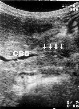
An 8-year-old girl from a low socio economic background was admitted with history of fever with chills and rigors of one-month duration, intermittent abdominal pain and bilious vomiting of 15 days duration. There was history of passing round worms in the stools and constipation. She was febrile, anemic, ill looking, toxic, dehydrated with icterus and had dry coated tongue. Pulse rate was 100/minute, and B.P 110/70 mmHg. The abdomen was tender with guarding, hyperesthesia and moderate hepatomegaly. She had no splenomegaly or ascites. Other systems were normal. Clinically, enteric fever was suspected. Investigations: Hemoglobin 7 g/dL; peripheral blood smear: normocytic hypochromic anemia, with no malarial parasite. Blood counts: TLC 4650/cumm, DLC: N - 72%, L -20%, E - 3%, ESR - 26 mm at first hr; blood Widal was positive with a titre of TO - l :240, T H - l :480, T AH - l :30 and T BH-l :30; Blood culture for Salmonella typhi was negative: Serum bilirubin 10 mg/dL (direct 7.8 and indirect 2.2 mg/dL); Serum AL T: 300 lU/L and serum alkaline phosphatase 350 lU/L. Sstool examination was normal. Ultrasound abdomen revealed a well distended gall bladder with wall thickness of 2mm and no evidence of calculi. The common bile duct was 6mm and was mildly dilated. There was a linear echo genic tubular structure with a central lumen within the distal CBD- suggests Biliary Ascariasis (Fig. 1).
The child was treated with intravenous fluids, antibiotics, and IV antispasmodics. She became asymptomatic after 48 hours. Subsequently, she was dewormed with a single dose of 400 mg of Albendazole. On follow up, a repeat stool and ultrasound abdominal examination were normal. The child had no recurrence of symptoms during the 8 weeks’ follow up. Discussion The biliary ascariasis may be complicated or uncomplicated(2). In uncomplicated biliary ascariasis the clinical picture merges with that of acute acalculous cholecystitis with low grade fever, upper abdominal colic, associated tenderness, muscle guarding in right upper quadrant with a gall bladder mass. Jaundice, hepatomegaly and marked toxemia are usually absent. The next common presentation is of acute cholangitis with high-grade fever, right hypochondriac pain, jaundice, tender hepatomegaly raised serum bilirubin, alkaline phosphatase and ALTs(1,2). The cholangitis may be suppurative in some patients and the patient may present with shock. We believe that our patient had acute non suppurative cholangitis. Ultrasound scan, CT scan, endoscopy and endoscopic retrograde cholangiography are methods employed in the diagnosis of biliary ascariasis. Plain X-ray films and ultrasound are found to be reliable, cost effective and widely available investigations for diagnosis and follow up of cases with biliary ascariasis(2,3). The worms are seen in ultrasound as linear or round areas of increased echogenicity representing worms, bull’s eye appearance and single or multiple long linear echogenic strips without acoustic shadowing within the bile duct. Real time scanning may reveal active movement of the worm within the biliary tract, which is diagnostic. Our case was diagnosed by ultrasound abdominal scan. With appropriate non-invasive management in the acute stage with IV-fluids and antispasmodics the worms will spontaneously return to the duodenum in 98% of the cases. Anti-helminthics are used after the acute stage has resolved to avoid killing or paralysing the worm in the biliary tract, as the dead worms may disintegrate and form a nidus for calculi. Complete recovery is usual in uncomplicated biliary ascariasis with a mortality rate of 1% or less(2). In conclusion, biliary ascariasis should always be considered as a probable diagnosis in a child presenting with fever, jaundice and severe abdominal pain. Non-invasive conservative management gives very good results. V. Harven Kumar,
|
![]()