|
Developmental vascular anomalies are common in neonates and infants. Portwine stains (culaneous hemangiomas) are always present at birth and may be a part of a syndrome. Klippel-Trenaunnay-Weber Syndrome is diagnosed by a triad of (a) Portwine nevus; (b) Hemihypertrophy; and (c) Venous varicosities.
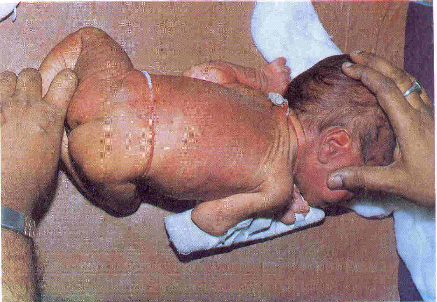
A 13-day-old second male child born of Hindu non-consanguineous parents presented with a portwine nevus spread all over face, scalp, back and left half of body
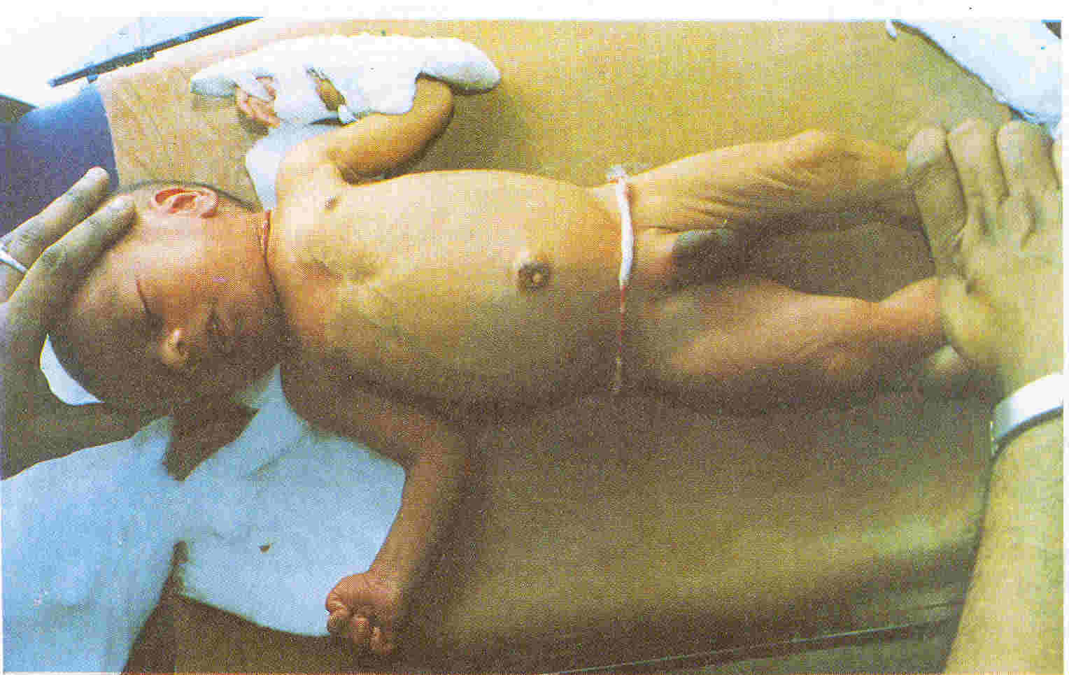
(upper and lower limbs) (Fig. 1) and venous varicosities over chest and abdomen (Fig.
2). There was left sided hemi-hypertrophy with the left thigh measuring 3 cm more and left arm measuring 1 cm more than the right sided counterparts (Fig. 2).
 |
|
Fig. 1. Extensive pottwine staining over face, trunk and limbs. |
 |
|
Fig. 2. Varicose veins extending over anterior abdominal and chest wall. Also seen is left sided hemihypertrophy (better seen over thighs). |
A normal platelet count excluded Kasabach-Merritt syndrome. CT Scan of brain and spine were normal, which ex- cluded Sturge-Weber syndrome (leptomeningeal Venous angioma, intracranial calcification,
etc.)
and Cobb Syndrome (spinal
arterio venous malformation). Rubinstein- Taybi syndrome was excluded by absence of characteristic facies and cardiovascular lesions. Normal Karyotyping excluded the
rare possibility of Trisomy-13 or Beckwith-Wiedman syndrome. Normal radiology of skeletal structures and absence of lymphangiectasis excluded Maffucci syndrome or Milroy's disease, respectively.
Soma De,
R.K. Jain,
Gautam Ghosh,
Shree Jain Hospital and Research Centre,
Howrah,
West Bengal,
India.
|
