|
|
Images in Clinical Practice Indian Pediatrics 2000;37: 907-908 |
|
Granuloma Annulare |
|
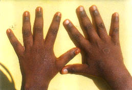
A 10-year-old male child was referred with history of asymptomatic skin lesions over the dorsa of hands, of nine months duration. He did not have any other cutaneous and/or systemic complaints. Physical examination revealed a few well defined skin colored papules typically arranged in an annular fashion giving rise to a ‘beaded’ appearance (Fig. 1). The lesions did not show any epidermal changes and the centre of lesion was hyperpigmented. Skin biopsy from the lesion showed focal degeneration of collagen surrounded by palisading histiocytes and fibroblasts (palisading granuloma). Granuloma annulare (GA) is a benign, usually self-limiting disorder occurring primarily in children. The most common type is the localized form with multiple lesions. It consists of small firm, asymptomatic papules that are skin coloured or erythematous and are often arranged in an annular or circinate fashion. The epidermal surface is most often undisturbed but sometimes it may show telengiectasiae. It usually occurs on the dorsa of the hands and feet. Unusual form of GA include: (i) Generalized form, consisting of hundreds of discrete or confluent papules usually seen over the trunk and proximal extremities; and (ii) Subcutaneous GA in which large painless deep dermal or subcutaneous nodules occur especially on palms, soles, legs, buttocks, fingers, eyelids and scalp. It may be confused with rheumatoid nodules, but can be differentiated by the absence of RA factor and arthritis; (iii) Perforating GA with umbilicatory lesions occurring on the hands and feet; and (iv) Eryhtematous GA in which the papular quality is less obvious than the erythema. Histo-logically all forms of GA share common histologic features namely focal degeneration of collagen in the upper and mid dermis with a histiocytic palisaded arrangement around the collagen bundles and abundant mucin. Granu-loma annulare may be associated with insulin dependent diabetes mellitus in children. Although, the disease is usually self limited, a variety of treatment modalities have been used to hasten the resolution process. These include topical corticosteroids or intralesional steroids (Triamcinolone), cryotherapy, laser and various systemic therapies, viz., PUVA, pentoxyfylline, nicotinamide, isotretinoin, salicylates, potas-sium iodide and dipyridamole.
G. Seturaman, Departments of Dermatology and |
|
|