|
|
Case Reports Indian Pediatrics 2000;37: 896-898 |
||
|
Dandy Walker Syndrome Associated with Unusual Congenital Anomalies |
||
From the Departments of Neurology and Neuro- surgery*, G.B. Pant Hospital, New Delhi 110 002, India. Reprint requests: Dr. M.M. Mehndiratta, Professor of Neurology, Department of Neurology, G.B. Pant Hospital, New Delhi 110 002, India. E-mail: [email protected]
Manuscript Received: November 10, 1999;
Dandy-Walker Syndrome (DWS) is defined as a congenital malformation of structures in the posterior fossa and is characterized by complete or partial absence of cerebellar vermis, cystic dilatation of fourth ventricle and frequently hydrocephalus. Clinical recognition has often rested upon radiological demons-tration of cyst like dilatation of the fourth ventricle and an anomaly of the cerebellar vermis. The etiology and pathogenesis of this condition is not yet determined but the presence of associated abnormalities is suggestive of an early embryonal developmental disturbance affecting more than one structure of the posterior fossa. A variety of central nervous system and systemic anomalies have been reported in association with DWS. A case of DWS with some unusual systemic anomalies (supernumerary nipples, cervical rib, pilonidal sinus, chordee penis and intrasacral meningo-cele), unreported in the literature so far, is being presented. The interesting thing is that teratogenic period of these anomalies coincides with DWS (5-8th week of embryonic period).
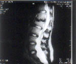
A 12-year-male reported to us with a history of delayed motor and mental milestones. He was born with low birth weight (1550 g) and had a delayed cry after birth. He had mild weakness in left upper and lower limbs since birth, which had increased in lower limbs for three to four years. In the last two months he had four episodes of generalized tonic-clonic seizures. Examination revealed dysmorphic face with lobulated pinna and low set ears, supernumerary nipples (right being more prominent than left), pigeon chest, thoracic kyphosis, pilonidal sinus and six cafe au lait spots (1.5 cm - 7 cm). Neurological examina-tion revealed mental retardation (Intelligent Quotient-65), cerebellar dysarthria, and spasticity of all four limbs (left more than right). The left plantar was extensor. Abdominal and cremastric reflexes were absent. The hemo-gram, routine blood chemistry, ECG, Ultra-sound of abdomen, liver, spleen, kidneys, echocardiogram, electromyography and nerve conduction velocity studies were normal. Radiological study revealed spina bifida at L4 vertebra and a left sided cervical rib. CT scan showed a posterior fossa cyst communicating with the fourth ventricle, hydrocephalus and cerebellar hypoplasia. MRI lumbosacral spine showed intrasacral meningocele with cord ending at L4 vertebrae causing tethered cord.
Although, DWS was described in the literature during the second half of the nineteenth century, it was only in 1914 that Dandy and Blackfan(1), published the first clinical description postulating at the same time the pathogenetic theory of absence of foramina of Luschka and Magendie. The abnormalities associated with DWS are either malformations of the brain or systemic anomalies(2). The case reported hereby is unusual in the sense that anomalies like supernumerary nipples, pilonidal sinus, chordee penis, cervical rib and intrasacral meningocele with tethered cord have not yet been reported in association with DWS(2,3). Interestingly the teratogenic period of some of these anomalies is also same as of DWS, i.e., 5th-8th week of embryonic period(4). The mammary gland and nipples develop from one of the mammary ridges at the sixth week of antenatal period. The cervical rib chondrifica-tion begins at 6th week of embryonic develop-ment. The chordee penis develops due to non-fusion of ventral urethra causing ventral fibrous band at 8th week of embryonic development. The pathogenesis of pilonidal sinus (congenital or acquired) is still contro-versial, but in this context, since it is associated with above mentioned congenital anomalies, it is likely to be of congenital origin. Intrasacral meningocele also occurs due to failure of closure of neural tube at 5-8th week of embryonic period(4). Thus all the above mentioned unusual unreported associated anomalies have the same teratogenic period (5th-8th) as of DWS except pilonidal sinus which has a controversial pathogenesis. | ||
|