|
|
Recommendations Indian Pediatrics 2000;37: 845-851 |
|||||||||||||||
|
Consensus Report on Neonatal Cholestasis Syndrome |
|||||||||||||||
|
E-mail: [email protected]
Neonatal Cholestasis Syndrome (NCS) includes a wide spectrum of clinical conditions ranging from congenital malformations of the hepatobiliary tree, infections, inborn errors of metabolism to some of the recently identified clinical conditions with or without genetic predilection. NCS has largely remained ignored in our country. There is scarcity of published (peer reviewed) data on NCS from India(1-5). The annual conference of Commonwealth Association of Pediatric Gastroenterology and Nutrition (CAPGAN) and Indian Academy of Pediatrics (IAP) Subspecialty Chapter on Pediatric Gastroenterology held in New Delhi in November 1998, in one of the scientific sessions entitled "Neonatal Cholestasis Syndrome: Indian Scene"(6) brought out a recommendation to have a consensus meeting with major objectives to adopt a uniform approach, give thrust on awareness and referral pattern and strengthen services of histo-pathology and laboratory. The national consensus meeting on the approach and management of NCS was organized on April 28th, 1999 at Sanjay Gandhi Postgraduate Institue, Lucknow. Data (experience and questionnaire) on prescribed proformas were obtained from eight medical centers (Annexure 1). Surgical data (experience and questionnaire) were obtained from six surgical centers (Annexure 1). Medical and surgical experience of all the centers were pooled and analyzed. The issue was deliberated upon during the meeting by a group of experts from different disciplines (Annexure 2).
The salient observations were: (A) NCS constitutes 30% of hepatobiliary disorders in India; (B) There is a long delay by parents in seeking medical attention for these infants (average duration 4.5 weeks); and (C) The average age of presentation to a specialized center is 3.5 months (range birth to 15 months) with a consequent delay of 3 months in referral (medical and surgical centers). Even after reporting to large referral hospitals, lack of requisite diagnostic facilities and inadequate expertise in handling such cases further adds to the delay which eventually deprives these infants of an opportunity of utilizing the current available therapeutic modalities. (D) Combined Experience Medical Of the total of 1008 analyzed cases of NCS, hepatocellular causes were seen in 53% (neonatal hepatitis 47%, metabolic 4% and 2% due to varied etiology), obstructive causes in 38% (biliary atresia 34%, choledochal cyst 4%), ductal paucity in 3% and other 6% cases were idiopathic. Among neonatal hepatitis (n = 468) idiopathic giant cell hepatitis constituted 64% and TORCH infections 22% (of these 22%, CMV was seen in 58%, toxoplasmosis in 23%, hepatitis B in 10%, rubella in 4.5%, syphilis in 4% and herpes in 1%), sepsis 8% and in 6% cases other causes like malaria, UTI, etc., were found. Among the metabolic group (n = 43) 35% were due to galactosemia, 33% due to AIAT deficiency, 19% had TPN related, 7% had tyrosinemia, 4% had storage disorders and 2% had hemochromatosis. The group of varied etiology (n = 22) due to hepatocellular causes were constituted by inspissated bile plug syndrome (n = 9), recurrent intrahepatic cholestasis (n = 2), progressive familial intrahepatic cholestasis (n = 1), hypothyroidism (n = 4), associated Down’s syndrome (n = 3); and one case each due to polycystic disease, postintestinal surgery and immunodeficiency. Ductal paucity (n = 29) was due to non-syndromic variety in 83% and syndromic variety in 17% cases. Data collected from medical centers revealed that 9% of cases subjected to laparotomy had normal ductal system. Surgical This is proforma based data acquisiton and analysis from six surgical centers (study period 1-15 years, total number of biliary atresia cases was 391). Diagnostic yield of prelaparotomy liver biopsy was higher in the experience of those groups who routinely perform a liver biopsy. Of the 358 cases subjected to surgical exploration, 78% had biliary atresia. Negative laparotomy for biliary atresia was higher (28%) in cases where prelaparotomy liver biopsy was not done vs 11% where prelaparotomy liver biopsy was done. Types of atresia were 90% type III, 7.8% type II and 1.7% type I. Cirrhosis was observed in 75-100% of cases at laparotomy, a reflection of the delay in diagnosis, referral and surgery. Surgical procedures of portoenterostomy were done in 86% (n = 242) and hepatico-jejunostomy in 3.2% (n = 9) cases. Perioperative mortality of average 6.7% (range 0-10%) was observed. Successful surgery was observed in 5-100% (mean 49%) cases with survival rates of 37.5%-100% (mean 56%) over a follow up period of 6 months. Jaundice free survivals were not analyzed. Marked variation in the results was observed on analysis of data from these centers. This is probably attributed to nonuniformity in definition, limitation of method of data acquisition and incomplete followup. Experts deliberated on medical, surgical and histopathology experience in India, problems faced in the country with regard to late referrals, lack of laboratory facilities for metabolic disorders, issues related to various diagnostic modalities including liver biopsy, radionucleide scan, lack of uniformity in diagnostic and therapeutic approach within the scanty number of specialized centers and concept of building a closely interlinked team of Pediatric Gastroenterologists/Pediatricians with special interest in Gastroenterology, Pediatric Surgeons and Pathologists. The group agreed on the following guidelines/recommendations.
Early referral of NCS infants can be ensured by educating the parents as well as different categories of health personnel who are consulted for treatment. Importance of jaundice in later neonatal period and possible consequences should be highlighted to create awareness in the community. There is a need to strengthen the available messages on early detection and referral to babies with NCS in Reproductive and Child Health Program. Possibility of having a parent information/education program can be explored. "Newborn babies having jaundice beyond 14 days of age with dark urine with or without acholic stools should be referred to appropriate health facility for further investigations and treatment without loss of time" should be a clear message to the health personnel who come across such cases. Diagnosis and management of NCS needs to be incorporated in under-graduate and postgraduate teaching curriculum in Pediatrics. Large/referral hospitals need to be sensitized and encouraged to investigate and treat cases with NCS. Early referral of babies with NCS is critical for the overall prognosis. Early surgical intervention results in better survival, particularly if cases are operated within 60 days. However, referring physicians/practitioners at all levels need to be educated that there is no cut off for late referral. Late portoenterostomy in those cases who report late can still be beneficial though with lower success rates and it offers a future scope for liver transplantation.
Babies with a clinical profile suggestive of NCS should be referred to the nearest hospital with facilities for investigating and treating such cases. It is essential to have a team of Pediatrician, Pathologist and Pediatric Surgeon in these specialized centers.
Cases with suspected extrahepatic biliary atresia (EHBA) should be referred to Pediatric Surgeon without loss of time. Even if liver biopsy is suggestive of neonatal hepatitis but the baby continues to have persistently pale stools, the patient should be referred to a Pediatric Surgeon. Surgical treatment of such cases should be contemplated in centers where at least 6 portoenterostomies are performed every year. Since the centers that fulfill this criteria are limited in our country, more and more surgical departments should be encouraged to develop proficiency in handling these cases.
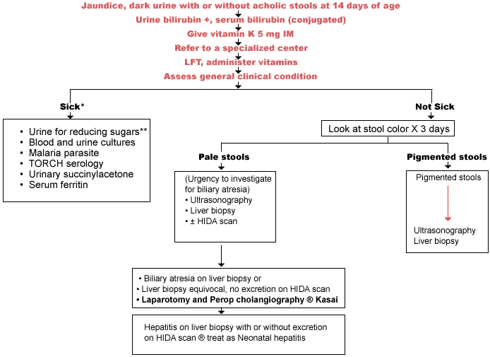
It is imperative that after hospitalization these babies are promptly investigated and a decision about the diagnosis and need for immediate surgical or medical intervention is taken without loss of time. The investigatory approach should take into consideration the clinical condition and presentation of the child (Fig. 1). In a sick baby with cholestatic jaundice possibilities of galactosemia, toxoplasmosis, herpes, tyrosinemia, sepsis, malaria, etc. should be considered. In babies apparently looking healthy and passing pale stools, serious consideration should be given to search for obstructive causes like EHBA or choledochal cyst on priority. These babies may have other causes like ductal paucity or even neonatal hepatitis. NCS cases who are passing pig-mented stools and do not look sick may be having neonatal hepatitis, ductal paucity, rarely metabolic/storage disorders or hypothyroidism; this group of children (passing pigmented stools) are unlikely to have biliary atresia. Work up of these cases should be based on a rational approach and all unnecessary investigations should be avoided. For instance, serum levels of alpha-1-antitrypsin (AIAT) are not very reliable to diagnose AIAT deficiency before 3 months of age. Therefore, in a suspected case it is useful to do phenotyping for AIAT deficiency or else wait and review after 3 months and perform deferred liver biopsy after 3 months of age for PAS staining. Liver biopsies of babies with suspected EHBA are an emergency and should be reported on priority basis. A uniform diagnostic criteria needs to be followed at all the centers. In case of any doubt about the histopathological diagnosis, the slides should be sent for second opinion to other centers with more experience without losing time.
The magnitude of metabolic causes appears to be underestimated in India because of lack of facilities for investigations. Since it is not possible to upgrade the laboratory services for each and every test at every place, it is cost effective to establish laboratory services for different tests for diagnosing metabolic dis-orders at one specialized center which serves as a referral center for others. Figure 1 - Approach to a Case of Neonatal Cholestasis Syndrome
There is a lot of variation in the manage-ment approach at different centers in the country which makes it difficult to share experience, validate the outcome at different centers and identify areas for research. A uniform management protocol (Fig. 1) is necessary for treating such cases and evaluating the outcome of various therapeutic interven-tions. In addition, parents and attending physi-cians need to be properly counseled regarding the expected outcome of the surgical inter-ventions. They should be told that the success of portoenterostomy can be judged by 6 months.
Cholestasis results in a variety of distressing symptoms and consequences of severe magni-tude which forms an important aspect of management of these cases. Nutritional support, vitamin supplementation and investigations at the earliest possible opportunity (Fig. 1) should be instituted. Those unfortunate babies who are not diagnosed on time consequently develop nutritional problems, pruritus, infection, portal hypertension, ascites and hepatic encephalo-pathy which need treatment. Nutritional
To the best our knolwedge injectable Vitamin E and water soluble formulations of vitamins A, D and K are not yet marketed in India. In the West these are available and are used. The above vitamin recommendations are based on current availability in India. Pruritus Pruritus is one of the distressing symptoms of NCS which can be reduced by giving phenobarbitone (5 mg/kg/day) alongwith UDCA (10-20 mg/kg/day). Cholestyramine (4-8 g/day) or rifampicin (10 mg/day) or photo-therapy with ultraviolet/infrared rays for 3-10 minutes in a day or naloxone and terfemadine (1-3 mg/kg/day) can also be tried to control pruritus. Infection Infection should be suspected in the presence of ascites and end-stage liver disease. Full work up for infection including ascitic fluid culture is necessary for appropriate antibiotic therapy. Cefotaxime should be used in peritonitis. If septicemia is suspected add amikacin or gentamicin. Associated Problems Portal hypertensions is always present in these cases. Variceal bleeding should be controlled with endoscopic sclerotherapy. Avoid use of NSAIDs. Ascites requires bed rest, salt restriction (Na + 1 mEq/kg/day-0.5 to 1 g salt). Fluids should be restricted to 70%. All bakery products and diets containing extra salt should be avoided. Diuretics like spirono-lactone and frusemide combination should be used. Intractable and tense ascites may require paracentesis. Hepatic encephalopathy should be treated with oral lactulose, oral amoxycillin or metronidazole, colonic washes, restricted proteins, evacuation of blood from GIT, correction of hypoglycemia, electrolyte imbalance and blood loss. Avoid renal failure as far as possible. Liver transplantation may remain the only option for infants with EHBA with decompen-sated liver disease (ascites and/or encephalo-pathy) or failed portoenterostomy.
The cooperation of all the medical and surgical centers for providing their data is acknowledged. Special thanks are due to Dr. S.R. Naik, Head, Department of Gastro-enterology, Sanjay Gandhi Postgraduate Institute of Medical Sciences (SGPGIMS), Lucknow and Dr. Anil Dhawan, Consultant, Pediatric Hepatology, Kings’ College, London, UK for their valuable academic inputs. The faculty members, senior residents and the staff of the Department of Gastroenterology, SGPGIMS, Lucknow deserve appreciation for organization of this meeting. Dr. A.K. Patwari and Dr. S.K. Yachha were responsible for recording, compilation of academic contents and preparing manuscript of consensus meeting. The administration of Sanjay Gandhi Postgraduate Institute of Medical Sciences, Lucknow provided infrastructure support and pharmaceutical companies provided financial support for organization of this meeting.
Medical: S.K. Mittal (Delhi), S.R. Naik (Lucknow), Anand Pandit (Pune), A.K. Patwari (Delhi), M. Sathiyasekaran (Chennai), B.R. Thapa (Chandigarh), A. Sibal (Delhi), M. Dwivedi (Allahabad), A. Bavdekar (Pune), U. Jhamb (Delhi), S. Gopalan (Delhi), G.K. Malik (Lucknow), Archana Kumar (Lucknow), A. Srivastava (Lucknow), A.K. Antony (Lucknow), Anil Dhawan (London), S.K. Yachha (Lucknow). Surgical: V. Bhatnagar (Delhi), S.J. Karmarkar (Mumbai), K.L. Narasimhan (Chandigarh), S. Shrotriya (Pune), S. Aggarwal (Delhi), S.S. Sikora (Lucknow), R. Saxena (Lucknow). Pathology: R.K. Gupta (Lucknow), V. Malhotra (Delhi), S. Datta Gupta (Delhi). | |||||||||||||||
|
|