A 4-year-old boy, presented with blistering and scarring of skin. He had
vesicles and erosions with mutilations of the skin over exposed surfaces
(Fig.1). There was hypertrichosis over face and extremities,
teeth were stained red (Fig. 2) with moderate splenomegaly. The
urine was red with increased levels of urinary and erythrocyte
porphyrins. A diagnosis of Congenital Erythropoeitic Porphyria was made.
 |
|
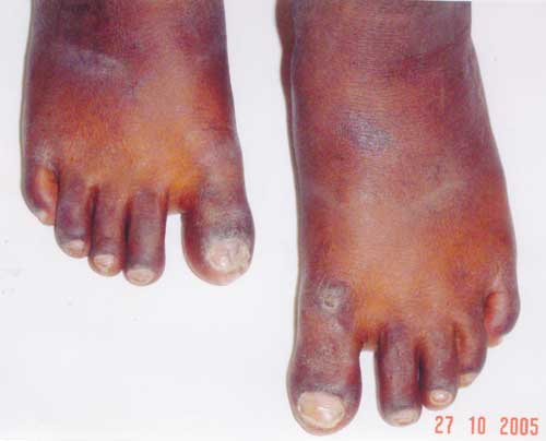
Fig. 1. Mutilated skin oner dorsum of first
toe (left), and tip of first toe (right) in porphyria.
|
 |
|
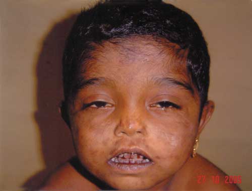
Fig. 2. Facial scarring, erosion, hyper
pigmentation and erythrodontia in a patient with congenital
erythropoeitic porphyria.
|
This autosomal recessive condition besides its
typical skin, dental and urine findings can also have ocular and
hematologic findings. Erythrodontia can also be seen in fluorosis,
tetracycline therapy, food stains or dentinogenesis imperfecta.
Porphyrial skin lesions must be differentiated from xeroderma
pigmentosum, epidermolysis bullosa and pemphigoid. The best therapy is
avoidance of sunlight, while oral beta-carotenes have been tried with
limited benefit.
A.N. Prasad,
Pediatrician,
Military Hospital,
Mhow, 453 441 (M.P.), India.
E-mail:
[email protected]
