From the Department of Radiology, Dr. Muhittin Ülker
Emergency Hospital, Ankara, Turkey; and *Department of Radiology, School
of Medicine, Fatih University, Emek, Ankara, Turkey.
Correspondence to: Asli Köktener, Department of
Radiology, School of Medicine Fatih University,
P.K. 5 Emek, 06510, Ankara/ Turkey. E-mail:
[email protected]
Manuscript received: September 1, 2004, Initial review
completed: October 29, 2004;
Revision accepted: February 21, 2005.
Abstract:
Sever’s disease (calcaneal apophysitis) is a self-limiting condition
seen in physically active children. Although there is controversy
about the radiographic appearance, some reports propose the importance
of fragmentation of the secondary nucleus in the diagnosis of Sever’s
disease. We studied secondary nucleus of the calcaneus with
ultrasonography. Twenty-one symptomatic heels of 14 children were
examined. All these heels showed fragmentation of the secondary
nucleus on both conventional radiograph and sonography.
Ultrasonographic examination also showed 2 retrocalcaneal bursitis.
Our initial data showed that sonography may be valuable in the
diagnosis of Sever’s disease.
Key words: Apophysitis, Calcaneus, Sever’s disease,
Ultrasonography
In 1907, Haglund described calcenodynia in
children and adolescents(1). In 1912, Sever reported this clinical
condition as osteo-chondrosis as ischemic changes of the secondary
nucleus(2). Similar to Osgood-Schlatter disease (OSD) (tibial
tuberosity apophysitis), Sever’s disease is an overuse syndrome
frequently seen in physically active children and adolescents between
the age of 8 and 10 years in girls and 10 and 12 years in boys(3,4).
The radiographic aspect of the secondary nucleus of
the calcaneus in children with heel pain remains controversial. The
recent studies stated that the fragmentation of the calcaneal
secondary nucleus was the most typical finding and also showed the
disappearance of the areas of disintegration after treatment of the
apophysitis(5,6).
In this study, we report our preliminary results
showing the ultrasonographic aspect of Sever’s disease and emphasizing
that sonography might be a diagnostic tool without radiation hazard to
the children.
Material and Methods
Twenty-one symptomatic heels of 14 children (2
girls, 12 boys) age 9-15 years (mean; 12.7) who were active in sports
were studied with ultrasonography because of the typical clinical
Sever’s disease. The ultrasound examination was performed with 10 MHz
linear transducer (Acuson EUB 6000). In addition, lateral radiographs
of the heel were taken for all children. Heels were evaluated for the
soft tissue changes and fragmentation of the secondary nucleus of the
calcaneus.
Results
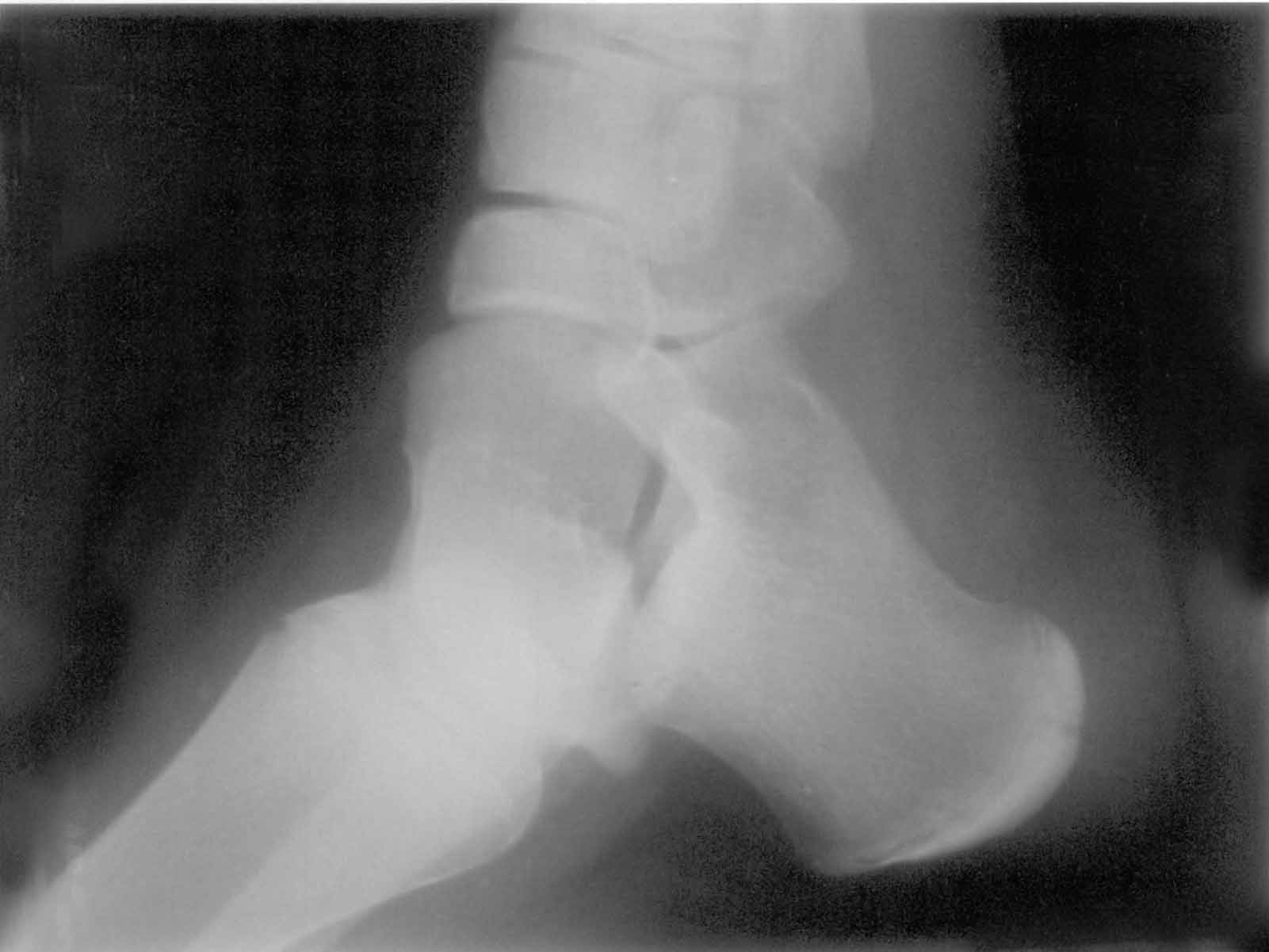
The radiographs of 21 symptomatic heels showed
fragmentation (Fig. 1). All of these heels (100%) had abnormal
sonographic examination showing the fragmentation of the secondary
nucleus (Fig. 2). Two of them also had retrocalceneal bursitis
demonstrated by ultrasonography. All 21 heels had normal achilles
tendon on sonographic examination. Only 3 heels were examined by
ultrasonography after one month’s treatment by the same person and
decreased fragmentation was observed.
 |
 |
Fig. 1. X-ray of the heel with fragmentation
of the secondary nucleus.
|
Fig. 2. Ultrasonography showing normal left
heel and right heel with fragmentation (arrows). |
Discussion
Sever’s disease, or apophysitis of the calcaneus is
a common cause of heel pain. The apophysis of the os calcis is an
epiphyseal plate that develops along the posterior border of the bone.
The achilles tendon inserts in the calcaneus apophysis. The growth
plate is weak and subject to injury. The most significant etiologic
factor in Sever’s disease is overuse and microtrauma in sports(7).
When Sever reported this condition as
osteochondrosis, sclerosis and fragmentation were demonstrated as
diagnostic X-ray findings. However, years later, there is still
controversy about the radiographic aspect of the calcaneal apophysitis.
Some authors showed that sclerotic changes could be observed in normal
children (7-9). Nery, et al. (1996) and Volpon, et al. (2002) stated
that fragmentation was the most reliable X-ray finding in calceneal
apophysitis(5,6). There have been studies including sonographic
features of the OSD that involves tibial tuberosity. Showing
pathologic findings like pretibial swelling, fragmentation of the
ossification center, insertional thickening of the patellar tendon and
excessive fluid collection in the infrapatellar bursa, they supported
the sonographic examination of knee as a simple and reliable method to
diagnose OSD(10-12). In Sever’s disease, ultrasonographic examination
provides to examine not only secondary nucleus of calcaneus but also,
Achilles tendon and retrocalcaneal bursa. Achilles tendinitis and/or
retrocalcaneal bursitis may accompany with Sever’s disease or may be
solely a cause of heel pain.
Our preliminary results showed that ultrasonography
could demonstrate the fragmentation of secondary nucleus of
ossification of the calcaneus and surrounding soft tissues. This
finding might be valuable in the easy diagnosis of Sever’s disease
since children are prevented from excess radiation. This study is the
first step, and further studies are needed to support the value of the
sonographic examination in the diagnosis of Sever’s disease.
Contributors: BH was involved in the examination of
the patients; AK did the literature search and prepared the
manuscript; GD reviewed the document.
Funding: Nil.
Competing interests: None.
