Rhabdomyosarcoma (RMS) is an aggressive malignant striated muscle
neoplasm and accounts for 50% of all pediatric soft tissue sarcomas.
Within the head and neck, common sites are orbit, paranasal sinuses
and soft tissue of cheek and neck. Paraoral RMS is rarely
reported(1,2). Tumors developing in a preexisting lesion are very
rare(3,4). Here we describe a case of Embryonal Rhabdomyosarcoma
arising from a congenital lesion involving the lip.
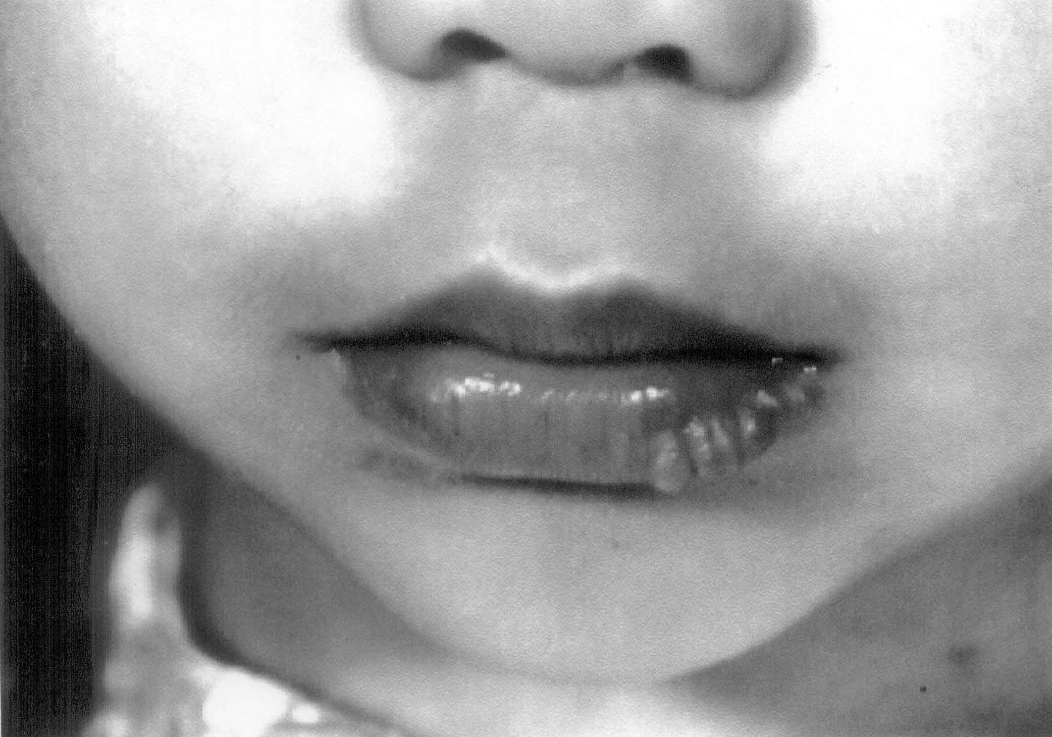
A 2½-year-old female child presented with history
of a lower lip swelling since birth. The lesion increased in size over
2 months. On physical examination a 1.5 × 1 cm lobulated lesion, with
multiple, small raised, irregular flesh colored rugae was present on
the lower lip. It was nontender, and nonerythematous (Fig. 1).
No lesions were present on the mucosal surface and the mass did not
cross the midline of face. Staging work up was done. Her hemogram,
biochemistry, MRI of head, face, orbit and brain, bone marrow and bone
scans were all normal.
 |
|
Fig.1. RMS of lower lip. |
The lesion was biopsied and histology reported as
Embryonal Rhabdomyosarcoma, intimately associated with small nerve
twigs. The tumor cells were strongly immunoreactive for desmin and
small areas positive for S-100 and neuron specific enolase.
The patient was operated and surgical margins were
free of tumor. She was started on adjuvant systemic chemotherapy
containing vincristine, ifosfamide, actino-mycin D (VAI). Current
therapy for RMS is vincristine, cyclophosphamide and actino-mycin D (VAC).
The patient was on a study, which showed other regimens to be
equipotent. The patient has completed chemotherapy and is in follow up
for last 5 years and is in remission.
This young child had a preexisting benign lesion of
the lower lip (cyst, hamartoma or amelanotic nevus) that transformed.
There have been two reports of RMS arising from a pre existing lesion,
the first, a case of a cutaneous melanotic nevus, and the second a
cystic adenomatoid malformation of the lung (3,4).
Immunohistochemically, this tumor was strongly
positive for desmin. S100, and neuron specific enolase (NSE) were
positive focally, an usual finding in RMS. Fetal rhabdomyoma, a benign
lesion found in the head and neck area of young children shows S 100
positivity. However the histology was not suggestive of rhabdomyoma.
However cutaneous RMS may show staining for neural elements and have a
predilection for the face(5).
We obtained excellent results in both survival and
cosmesis. The knowledge that benign cutaneous lesions may transform
into RMS will help in early diagnosis and satisfactory treatment for
any other affected children.
Tulika Seth,
Pamela Kempert,
Department of Medical Oncology,
Institute Rotary Cancer Hospital,
All India Institute of Medical Sciences,
Ansari Nagar, New Delhi 110 029, India.
E-mail: [email protected]
and
Department of Pediatrics Hematology Oncology
University of California, Irvine,
Irvine, California, USA.
