|
|
Brief Reports Indian Pediatrics 2004;41:817-821 |
||||||||||||||||||||||||||||||||||||||||||||||
|
Anterior Lens Capsule Vascularity in Evaluating Gestation in Small for Gestation Neonates |
||||||||||||||||||||||||||||||||||||||||||||||
|
Jitender Nagpal, Ajay Kumar and Siddharth Ramji From the Department of Neonatology, Maulana Azad Medical College, New Delhi 110 002, India. Correspondence to: Dr. Ajay Kumar, Associate Professor Pediatrics, Department of Neonatology, Maulana Azad Medical College, New Delhi 110 002, India. E-mail: [email protected] Manuscript received: July 18, 2003, Initial review
completed: September 12, 2003;
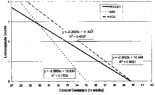
2 = 0.75) was stronger compared to SGA neonates (r2 = 0.50). Gestational age assessment by lens capsule grading had a better agreement with clinical gestation in AGA subjects (Kappa Index - 0.56 in AGA vs. 0.26 in SGA). The lens capsule grading under-estimated the gestation of SGA subjects by an average of three weeks compared to AGA neonates. Low birth weight is an important public health problem in the developing world. A large proportion of these babies, including preterms, are intrauterine growth retarded. A reliable estimate of the gestational age of such newborns is compounded by the non-availability of accurate menstrual dates and/or early pregnancy ultrasonographic examination. Therefore, alternative methods such as clinical scores and ophthalmoscopic estimation of the anterior lens capsule vascularity have been used in such cases. Hittner, et al.(1) described a simple method, based on the normal embryological process of gradual disappearance of the anterior lens capsule (ALC) vascularity between the 27th and the 34th weeks of gestation. ALC vascularity was categorized into 4 grades (4 = ALC vascularity covers entire anterior lens surface; 3 = early vascular atrophy with central clearing; 2 = more clearing with thinning of peripheral vessels; 1 = few peripheral thin vessels with none reaching center). These grades corresponded to 27-28, 29-30, 31-32 and 33-34 weeks of gestation respectively. A few studies have reported that intrauterine growth retardation has no effect on the disappearance of the ALC vascularity(2,3) while some case reports have suggested otherwise(4,5). This could be of significance in countries with a high prevalence of fetal growth retardation. The present study was designed to evaluate the effect of fetal growth retardation on the regression of the anterior lens capsule vascularity in low birth weight neonates. Subjects and Methods This prospective observational study was conducted in a tertiary level neonatal unit of a teaching hospital over seven months period starting May 2002. All intramural neonates with birth weights < 2000 g were recruited into the study. Those with major congenital malformations, including corneal opacity or cataract and evidence of TORCH infection were excluded. The data recorded in all neonates included history of maternal illness (diabetes, PIH/pre-eclampsia and chronic renal disease), evidence of perinatal asphyxia (apgar score, fetal bradycardia and/or meconium stained liquor), anthropometric parameters (birth weight, length and head circumference), gestational age (by LMP and first trimester ultrasonography) and intrauterine growth status(6). In all subjects the gestational age was clinically assessed using Ballard’s Score(7) by one of the investigators (JN) within 24 hours of birth. The gestational age assigned to the subjects was based on the following criteria: (a) If date by the last menstrual period (LMP) or first trimester ultrasound, and clinical assessment were within two weeks, then LMP derived age was the assigned gestation; (b) If LMP was uncertain or the difference was more than two weeks between the LMP (or first trimester ultrasound) and Ballard score, then gestation by Ballard’s score was the assigned gestation. The direct ophthalmoscopic evaluation was done by either of the two investigators (AK, SR) who were blinded to the gestation of the neonates within 24 hours of birth after full dilatation of the pupils using tropicamide 0.5% and 2.5% phenylephrine. The vascularity of the anterior lens capsule was classified in accordance with Hittner’s grading(1). The data was analyzed using Epi-Info 2002 software. Groups were compared using Chi-square, Fisher-exact and Kappa statistic. Linear regression analysis was used for correlation between gestational age and ALC vascularity grade. Results There were 157 eligible neonates during the study period. Eight neonates were excluded (7 with major congenital mal-formation and 1 with TORCH infection). Ten neonates could not be evaluated, as they were critically sick and expired within eight hours of birth. Remaining one hundred and thirty nine neonates of 27-40 weeks gestation were finally included in the study (107 preterm and 32 full term). Table 1 depicts baseline clinical variables between the growth retarded (SGA) and non-growth retarded (AGA) neonates. The two groups were comparable for all variables, except gestation, which was significantly higher in SGA compared to AGA neonates. Table 1 also provides the comparison for ALC vascularity and clinical gestational age agreement in SGA and AGA neonates. The kappa statistics for agreement was 0.56 in AGA neonates and 0.26 in SGA neonates.TABLE I Baseline Characteristics of SGA vs. AGA Babies.
* p < 0.0001 Correlation between the grades of the lens capsule vascularity and gestation is depicted in Fig. 1. In 20% of the enrolled neonates the lens capsule vascularity persisted well beyond 34 weeks (27/79 in SGA vs. 1/60 in AGA) and the correlation for the cohort unadjusted for intrauterine growth status was poor (r2 = 0.45). The correlation in AGA neonates was better (r2 = 0.75) as compared to SGA neonates (r2 = 0.50). A plot of the gestational age difference between the two assessment methods for SGA and AGA babies revealed that lens capsule vascularity underestimated the gestational age in SGA neonates on an average by about 3 weeks (range 1-5 weeks) compared to the AGA neonates.
Discussion The examination of lens vascularity in AGA neonates has been consistently found to have a good correlation with clinical estimates of gestational age(8,9) and Hittner’s grading has been accepted as a valid method of gestational age assessment in such neonates. In small for gestational age babies also a similarly good correlation has been demonstrated by a few workers(2,3). The impact of intrauterine growth retardation on the rate of regression of the anterior lens capsules vascularity however continues to be unresolved. Hittner, et al. in 1981(10) conducted a study on 33 small for gestational age infants and found a significant negative correlation between gestational age and the grade of the anterior lens capsule (r = – 0.77). However in one third of the subjects the vascularity of the lens capsule persisted beyond 34 weeks. These observations are similar to the present study (r = – 0.71; r2 = 0.5031) including 34 percent of the SGA infants having persistence of vascularity beyond 34 weeks. Hittner, et al.(1) in a previous study on AGA neonates had demonstrated the correlation coefficient of –0.87 (r2 = 0.76) which is comparable to the findings of the present study (r = –0.86; r2 = 0.75). Anecdotal case reports(4,10) also suggest that there is a delay in the regression of the ALC vascularity in SGA neonates. Two earlier studies(2,3) designed to study the effect of various perinatal factors on the regression of ALC vascularity have concluded that intrauterine growth retardation had no significant effect on the relationship between the lens capsule grade and the clinical gestation suggesting that the grading was equally valid in the presence of such factors. However these studies have been conducted using small sample size in settings where the prevalence and severity of intrauterine growth retardation are likely to have been low. A larger sample size, increased severity of intrauterine growth retardation and greater prevalence of growth retardation caused by nutritional deprivation are likely to have been the reasons for the emergence of a statistically significant difference between the SGA and AGA babies in the present study. No data is currently available as to the possible reasons for the delay in the regression of the anterior lens capsule vascularity in SGA babies. However it may be hypothesized that since the vascular system serves to nourish the growing lens, in nutritionally deprived fetuses such nourishment may be required for a longer period for the complete growth of the lens allowing for the persistence of the vascular structures in such a situation. The present data supports the earlier observations that the regression of the anterior lens capsules has a negative correlation with gestation. However, in the case of SGA neonates the correlation is weaker and the regression appears to continue up to 39-40 weeks. Thus, in regions of the world wherein the prevalence of intrauterine growth retardation is high, especially even amongst preterm neonates, the utility of anterior lens capsule vascularity for gestation assessment is limited. Acknowledgements The authors would like to acknowledge the contribution of Dr. Jyoti Dhaka and Dr. Puneet Kochar who were involved in the recording of baseline information of the subjects. Contributors: All the three authors were involved in planning and designing the study. JN carried out the Ballard’s assessment and did the statistical analysis and drafted the manuscript. AK and SR did the ophthalmic examination, helped in the analysis and critically reviewed the manuscript. SR shall act as guarantor for the paper. Funding: None. Competing interests: None.
| ||||||||||||||||||||||||||||||||||||||||||||||
|
References | ||||||||||||||||||||||||||||||||||||||||||||||
| ||||||||||||||||||||||||||||||||||||||||||||||
![]()