|
|
Case Reports Indian Pediatrics 2002; 39:761-763 |
||
|
Cerebro-Oculo-Facio-Skeletal Syndrome in a Neonate |
||
N.B. Mathur
R.K. Alwadhi
Pankaj Garg
Cerebro-oculo-facio-skeletal (COFS) syndrome was described by Pena and Shokeir as a combination of severe microcephaly and ocular abnormalities with characteristic facies comprising of large pinnae, blepharophimosis, prominent root of nose, upper lip overhanging lower one, long philtrum and micrognathia(1). Other important features include contractures at elbow and knee, camptodactyly and rocker bottom feet. Failure to thrive associated with marked hypotonia and developmental retardation was also consistently seen in the ten cases described in the initial report. Subsequently twenty more cases have been reported in literature till date(1-7). We are reporting one such case. Case Report A 7-day-old full term male baby born out of non-consanguineous marriage presented to our nursery with complaints of not opening eyes. Antenatal period was uneventful. Baby was delivered at home, was active and exclusively breastfed.
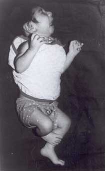
At presentation the anthropometry revealed weight 2.33 kg; length 46.5 cm and head circumference 26.5 cm. The infant had microcephaly with facial dysmorphism (Fig.1) Anterior fontanelle was 0.3 × 0.3 cm, coronal and lambdoid sutures were ridged. Examination of the eye revealed microphthalmia, blepharophimosis and bilateral central corneal opacity. Ears were large, root of nose was prominent, upper lip was overhanging the lower one and micrognathia was present. Hands revealed bilateral fixed flexion deformity at proximal interphalangeal joint of little fingers. Systemic examination was within normal limits. On investigation CT cranium showed craniosynostosis with coronal and lambdoid sutures showing premature fusion and cerebral atrophy. Abdominal USG, karyotype and echocardiogram were normal. Serology for rubella was negative. Infantogram showed normal mineralization of bones. The child was discharged after 7 days on same weight; returned with diarrhea after 2 weeks with weight loss. Subsequently he was discharged on request after the loose stools settled. Discussion COFS syndrome is characterized by microcephaly, microphthalmia and characteristic facies. Causes of microphthalmia and microcephaly are given in Table I. The more common syndromes like Rubella, Trisomy 13 and Trisomy 18 can be excluded by the absence of their hallmarks clinically and subsequently on negative serology for rubella and a normal karyotype. Other less common syndromes with microcephaly and microphthalmia are Meckel Gruber, Hallermann Streiff, Fanconi pancytopenia and Goltz. Microphthalmia is occasionally seen in Goltz. The deletion syndromes can also be excluded clinically and on karyotyping. Table I- Causes of Microcephaly with Microphthalmia. Rubella Syndrome Trisomy 13 syndrome Trisomy 18 syndrome Cockayne syndrome COFS syndrome Meckel Gruber syndrome Hallermann Streiff syndrome Fanconi pancytopenia syndrome Goltz syndrome 4p - syndrome 13q - syndrome 18q - syndrome
Cockayne syndrome (CS) is closely related to COFS syndrome. Neuropathological changes of microencephaly, enlarged ventricles and patchy or discontinuous demyelination are common to both COFS(8) and CS. Features in the present report which are common to COFS and CS include microcephaly, microphthalmia, corneal opacities, blepharophimosis and large ear pinnae. Features favoring COFS include presence of camptodactyly, which is not observed in CS. Further, microcephaly is not congenital in CS. Cryptorchidism and sparse hair have been observed in CS. Isolated case reports point to possible rare associations like congenital muscular dystrophy like changes(9), intracranial calcification(10) and accelerated cochlear nerve degeneration(11). Out of thirty cases reported in literature, 20 are from Manitoba, Canada. Evidence strongly suggestive of autosomal recessive inheritance has been observed(1). Increasing awareness and recognition of this syndrome is exemplified by a recent report of prenatal diagnosis of this syndrome using transabdominal USG on 21-week-old fetus. Features seen included severe bilateral microphthalmia, micrognathia, multiple joint contractures and rocker bottom feet(12). Contributors: NBM was involved in analysis of findings and finalization of draft. RKA and PG were involved in drafting of the manuscript and primary care of the patient. Funding: None. Competing interests: None stated.
| ||
|
References | ||
|
![]()