The management of pyothorax and pneumothorax
involves diagnostic or therapeutic aspiration and intercostal drainage
(ICD) tube insertion. These procedures are considered minor and done
under local anaesthesia, usually without sedation in children. Rarely
these procedures may get complicated with some human- or
instrument-related mishaps. Two such cases of empyema with unusual
iatrogenic foreign bodies are presented here. The role of thoracoscopy
in removal of such pleural foreign bodies is highlighted.
Case Reports
Case 1. A 21-day-old girl presented with
cough for 15 days, rapid breathing and high grade fever for 2 days.
Clinical examination and chest X-ray revealed the presence of
right sided pyopneumothorax. Supportive treatment for management of
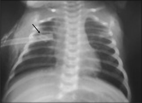
septic shock started and urgent ICD tube insertion using a Malecot’s
catheter were tried. When manipulating for insertion, a part of the
Malecot’s catheter broke and got retained inside the pleural cavity.
Another Malecot’s catheter was inserted for drainage of pyopneumothorax
(Fig. 1). Thoracoscopic removal of foreign body was
planned after hemodynamic stabilization of patient.
 |
|
Fig. 1 Chest X-ray (PA-view)
showing Malecot’s catheter in right pleural cavity to drain
pyopneumothorax. Note the broken tip of the Malecot’s catheter
inserted at first attempt (arrow).
|
After three days, when the infant recovered from
septic shock and her lungs expanded significantly, she was operated
upon. Surgery was performed under general anaesthesia in left lateral
thoracotomy position. The foreign body was localized thoracoscopically.
But as patient was not maintaining oxygen saturation with carbon dioxide
pneumothorax required for procedure to continue, further port placement
was abandoned. The ICD site incision was enlarged to about 5 cm and
foreign body extracted under direct vision. Localization of foreign body
using thoracoscopy helped in its extraction with a smaller than usual
incision. Pleural cavity was lavaged and cleaned thoroughly. An ICD was
placed again. The infant recovered well and was discharged.
Case 2. A 2-year old male child, presented
with fever and cough for 2 weeks. Clinical examination was suggestive of
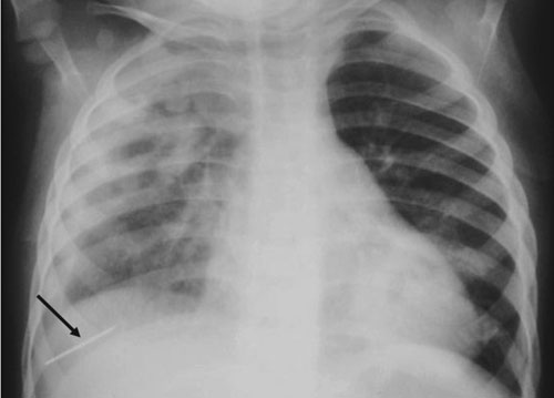
severe pneumonia with right-sided pleural effusion. His X-ray
chest showed patchy opacities in the right wing with effusion and left
lung hyperinflation. During aspiration of pleural effusion, the needle
got detached from its hub and got sucked into the pleural cavity (Fig. 2).
 |
|
Fig.2 Chest X-ray (PA-view)
showing needle in right costophrenic angle (arrow) broken while
attempting aspiration of pleural effusion.
|
Urgent thoracoscopy was planned in view of the sharp
foreign body. The child was placed in left lateral position and three
ports were placed: 1st in 6th intercostal space in posterior axillary
line and 2nd and 3rd in anterior axilla two spaces above and below the
first port, respectively. Needle was found lying in cardio-phrenic
angle. It was held with forceps and extracted along with port. Thoracic
cavity was lavaged thoroughly and ICD was placed. Patient recovered
well.
Discussion
Thoracic foreign bodies had broadly been divided into
three categories: (1) Aspirated Foreign bodies, which are most common
and involve tracheobronchial tree, (2) traumatic or accidental foreign
bodies which are more common in thoracic cavity; bullet being the most
common among this category and (3) Iatrogenic foreign bodies, which
occur as result of human and equipment error [1]. Among the iatrogenic
category, earlier the accidentally left gauze pieces and instruments
during a surgical procedures were the most frequent. These type of
foreign bodies are now reduced by 80% as a result of universal practice
of counting them before closure, and efforts are on to reduce it to
"never occurring event" [2].
Other form of pleural thoracic bodies are extremely
rare. On search of English literature we could find published reports of
retained pieces of ICDs [3,4], surgical blade which got detached from
scalpel [5], retained washer of rib approximator [6], broken blade of
foreign body forceps [1] and Abram’s needle [7], but all in adults.
These kind of iatrogenic complications can have predictable
consequences. Similar complications in future can be avoided by creating
increased awareness of such occurrence among doctors in training. As
such mishaps occur with struggling child leading to breakage of
instruments, it is suggested that proper stabilization of the patient by
using sedation and restraints, where necessary, be used to avoid such
occurrences. Moreover, removal of such a foreign body should be
undertaken after adequate hemodynamic stabilization of the child.
Thoracoscopy proved to be very useful procedure in
both these cases. In the first case, it helped in localisation of
foreign body and thus in retrieval through mini-thoracotomy incision. In
the second case, the removal of a sharp foreign body could be achieved
successfully using the thoracoscope. In both cases the procedure also
helped in clearing the purulent secretions and the debris due to empyema.
Thoracoscopy is gaining wide acceptability in removal of all types of
thoracic foreign bodies. The expected advantages of thoracoscopy over
thoracotomy in removal of FB are reduced pain, reduced chest wall
deformity (including winging of scapula and scoliosis), better
visualization and cosmesis.
Procedures for the management of empyema should be
done with utmost care and adequate sedation should be used to make the
patient comfortable. If these complications do happen, use of
thoracoscopy facilitates the management of any such condition and aid in
speedy recovery of patients.
1. Weissberg D, Weissberg-Kasav D. Foreign bodies in
pleura and chest wall. Ann Thorac Surg. 2008;86:958-61.
2. Regenbogen SE, Greenberg CC, Resch SC, Kollengode
A, Cima RR, Zinner MJ, et al. Prevention of retained surgical
sponges: A decision-analytic model predicting relative
cost-effectiveness. Surgery. 2009;145:527-35.
3. Paddle A, Elahi M, Newcomb A. Retained foreign
body following pleural drainage with a small-bore catheter. Gen Thorac
Cardiovasc Surg. 2010;58:42-4.
4. Gaucher A, Levrat Q, Troitzky A, Corbi P, Debaene
B, Mimoz O. Broken chest tube into the pleural cavity by a Monod’ trocar.
Ann Fr Anesth Reanim. 2010;29:153-5.
5. Singhal S, Dureja J, Kad N, Thakur A. An unusual
foreign body in the pleural cavity – an iatrogenic complication. Indian
J Thorac Cardiovasc Surg. 2010;26:233-4.
6. Abid Q, Devbhandari M, Davies H, Carr M. Missing
washer of the rib approximator? An easily overlooked foreign body.
Interact Cardiovasc Thorac Surg. 2003;2:108-10.
7. Fite E, Force L, Casarramona F, Verdaguer A.
Breakage and detachment of an Abrams needle in the pleural cavity during
performance of a pleural biopsy. Chest. 1989;95:928-9.


