|
|
|
Indian Pediatr 2015;52:
337-338 |
 |
Chromhidrosis – Colored Sweat in a Toddler
|
|
S Balasubramanian, Sumanth Amperayani, K Dhanalakshmi
and *Ram Kumar
From Departments of Pediatrics and *Dermatology,
Kanchi Kamakoti CHILDS Trust Hospital and The CHILDS Trust Medical
Research Foundation, Chennai, India.
Correspondence to: Dr S Balasubramanian, Senior
Consultant Pediatrician, Kanchi Kamakoti CHILDS Trust Hospital and The
CHILDS Trust Medical Research Foundation,12-A, Nageswara Road,
Nuangambakkam, Chennai 600 034, Tamil Nadu, India.
Email: [email protected]
Received: August 20, 2014;
Initial review: September 15, 2014;
Accepted: January 09, 2015.
|
|
Background: Chromhidrosis means
production of coloured sweat. Case characteristics: A toddler who
presented with colored sweat was diagnosed to have chromhidrosis based
on skin biopsy. No treatment was attempted considering the young age.
Outcome: Parents were counselled about the benign nature of this
disorder. Message: Identification of causes of colored sweat
requires appropriate investigations.
Keywords: Child, Chromhidrosis, Sweat glands.
|
|
C
hromhidrosis is the production of colored sweat
by eccrine or apocrine sweat glands. There have been very few reports of
chromhidrosis in the pediatric age group. We report a 3½-year-old girl
with this rare dermatological disorder.
Case Report
A 3½-year-old girl, the only child of
non-consanguineous parents presented with frequent bluish discoloration
of pillow covers after overnight sleep, noted by parents for last one
month. This was more obvious after sleeping in the day time in summer in
a non-air-conditioned bedroom. Her hat which was used by her during a
recent summer holiday was also noted to stain blue. There was no history
of drug intake or use of any topical coloring agents for the skin or
hair. Her urine and stools were normal. Clinical examination was
completely normal, and she did not have any specific odor.
Investigations suggested mild anemia (hemoglobin 10 g/dL), normal renal
and liver function, and normal metabolic screen. Skin biopsy from scalp
was sent for histopathological examination (HPE) with a specific request
to look for the presence of lipofuscin granules using periodic
acid-Schiff (PAS) staining. HPE was suggestive of Chromhidrosis and
parents were counselled about benign nature of this condition. We did
not consider any topical medication because of the young age, and also
because she was completely asymptomatic otherwise.
Discussion
Chromhidrosis refers to secretion of colored sweat
and was first reported in 1709 by Yonge of Plymouth [1]. It has been
classified into apocrine, pseudo-eccrine, and true eccrine chromhidrosis
[2]. Apocrine chromhidrosis is production of brown, black, blue, green,
or yellow colored sweat seen in axilla, face, and areolar region. It is
postulated to be occurring due to oxidized lipofuscins. These show
auto-fluorescence at 360 nm on skin and stained clothes, and are also
detectable by using auto fluorescence microscope on skin biopsy specimen
[3]. Pseudo-eccrine chromhidrosis is production of colorless sweat that
becomes colored when it reaches the skin and reacts with agents such as
chromogenic bacterial products, chemicals, paints, or dyes [4]. True
eccrine chromhidrosis is a very rare condition, occurring through
eccrine excretion of water-soluble agents like dyes and drugs [2]. It is
not associated with any known systemic disorders.
The disorder is chronic and may slowly regress as the
secretion of apocrine glands decreases with age [5]. Our patient
represents the second youngest reported case of apocrine
chromhidrosis, the youngest so far reported from Turkey [6].
Apocrine chromhidrosis is postulated to result from an increased
production of tyrosine, heme and melanin [7]. Color of sweat varies with
the oxidation status of the lipofuscin granules and may vary from
yellow, green, blue, brown, to black. Higher states of oxidation result
in a darker color [8].
 |
|
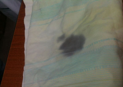
Fig. 1 Pillow cover with dark stain of
the patient’s sweat. (See color image at website).
|
Diagnosis is confirmed by increased number of
lipofuscin granules within chromhidrotic apocrine cells [6]. Emotional
or physical excitation may precede the onset of colored sweat.
Differentials include hyperbilirubinemia, Pseudomonas infection,
bleeding diathesis (red sweat, hematohidrosis), alkaptonuria (ochronosis),
and poisoning [8]. Successful treatment of apocrine chromhidrosis with
capsaicin cream 0.025% (mainly due to its topical counter-irritant
properties) has been reported. Relapse can occur within a few days of
discontinuing the medication [9]. There have been a few reports of
botulinum toxin being used for this condition.
Contributors: SB: diagnosed and managed the case;
SA, DK: literature search and prepared the manuscript; RK: skin biopsy
and histopathological examination.
Funding: None; Competing interest: None
stated.
References
1. Shelley WB, Hurley HJ. Localized chromhidrosis: A
survey. Arch Dermatol. 1954; 69:449-71.
2. Cilliers J, de Beer C. The case of the red
lingerie-Chromhidrosis revisited. Dermatol. 1999;199:149-52.
3. Barankin B, Alanen K, Ting PT, Sapijazko MJ.
Bilateral facial apocrine chromhidrosis. J Drugs Dermatol. 2004;3:184-6.
4. Leite RM, Nery NS. Dermatitis simulata: The
mystery of the blue girl. Int J Dermatol. 2007;46:1317-9.
5. Daoud MS, Dicken CH. Apocrine chromhidrosis. In:
Freedberg IM, Eisen AZ, Wolff K, Austen FK, Goldsmith LA, Katz SI,
Fitzpatrick TB, eds. Dermatology in General Medicine. 5th ed. New
York, NY: McGraw-Hill; 1999:811-2.
6. Carman KB, Aydogdu SD, Sabuncu I, Yarar C, Yakut
A, Oztelcan B. Infant with chromhidrosis. Pediatr Int. 2011;53:283-4.
7. Timani SS, Rubeiz N. Chromhidrosis. Available
from: www.emedicine.com/derm/topic596.htm. Accessed October 6,
2013.
8. Griffith JR. Isolated areolar apocrine
chromhidrosis. pediatrics. 2005;115;e239.
9. Marks JG. Treatment of apocrine chromhidrosis with topical
capsaicin. J Am Acad Dermatol. 1989;21:418-20.
|
|
|
 |
|
