|
|
|
Indian Pediatr 2012;49: 341 |
 |
Linear Atrophic Lesion on Forehead
|
|
Abhijeet Kumar Jha, Piyush Kumar and Sambeet Kumar Mallik
Department of Dermatology, Katihar Medical College
and Hospital, Katihar, Bihar, India.
Email: [email protected]
|
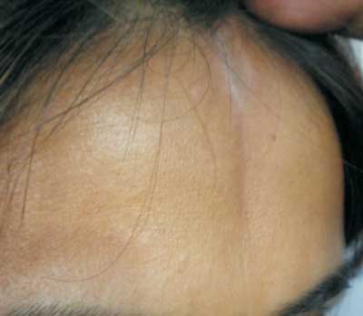
A 17 year old girl presented with asymptomatic linear
depressed groove involving the midline of the forehead
region which was progressively increasing over the past 1
year. The lesion was slowly extending to the scalp which
became a major concern to the patient. There was no history
of trauma preceding the development of the lesion. On
examination, an ivory sclerotic depressed linear lesion
mainly involving the mid forehead with slight extension to
the scalp was noted (Fig. 1). No other
remarkable cutaneous or systemic finding was noted. We
considered a differential diagnosis of "en coup de sabre"
morphea, and linear atrophoderma. On histopathology,
squared-off edge of the biopsy specimen with mild
superficial and deep inflammatory infiltrate with collagen
fibres, so a diagnosis of "en coup de sabre" morphea was
made.
 |
|
Fig. 1 Linear atrophic
lesion on forehead.
|
The name en coup de sabre is a french
phrase and came from its resemblance to sabre cut. It is a
localised type of linear scleroderma. The lesion usually
starts with contraction and firmness of the skin over the
affected area. Subsequently, an ivory irregular sclerotic
plaque develops, sometimes with telangiectatic vessels
coursing over it, together with hyperpigmentation at the
edge. It may involve the scalp, producing a linear zone of
alopecia, which may be preceded by bleaching of the hair.
The groove may extend downwards into the cheek, nose and
upper lip, and involve the mouth and gum. Subcutaneous
tissue, muscle, and, occasionally, bone are involved; this
ipsilateral form is known as progressive facial hemiatrophy
or Parry-Romberg syndrome. Severely affected patients may
have neurological manifestations due to involvement of the
meninges resulting in seizures, so it is very important to
diagnose and treat this condition at an early stage to avoid
complications. Most cases of linear scleroderma are
self-limited, with clinical activity apparent for an average
of 3 to 5 years but en coup de sabre may have an insidious
course lasting for decades. Treatment mainly is directed
towards the inflammatory component and consists of
corticosteroids, vitamin D analogues, methotrexate,
cyclophosphamide, azathioprine, hydroxychloroquine,
intralesional interferon- a,
D-penicillamine and psoralen with ultraviolet A therapy
(PUVA).
|
|
|
 |
|
