|
|
|
Indian Pediatr 2010;47: 346-347 |
 |
Cutaneovisceral Angiomatosis |
|
Saliha Senel and Nilgun Erkek,
Department of Pediatrics, Dr. Sami Ulus Children’s Health
and Diseases Training and Research Hospital, Ankara, Turkey. Email:
[email protected]
|
|
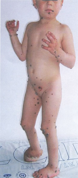
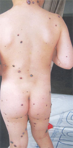
A 4 year old girl presented with painful hemorrhagic lesions all over her
body that had gradually increased in size and number since birth. Physical
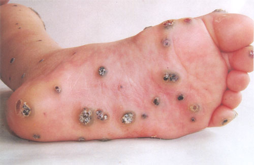
examination revealed multiple, various sized lesions localized on her
scalp, face, ears, lips, oral cavity, trunk, arms, palms, genital area,
legs and feet (Figs. 1 to 3). Her right foot had been
amputated in another center at the age of 2 because of multiple giant
hemorrhagic hemangiomas. Hemoglobin was 9.3 g/dL and platelet count was
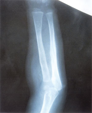
197x109/ mm3. X-ray
revealed radiolucent lesion on the diaphysis of the left ulna with
multiple small round calcifications in the surrounding soft tissues
interpreted as nonspecific lesions (Fig. 4). Abdominal
ultrasound and cerebral computed tomography scans were normal.
|
 |
 |
|
Fig.1 Frontal view showing multiple,
various sized lesions localized on face, ears, lips, trunk, arms,
palms, genital area, legs and feet.
|
Fig. 2 Back view of the patient showing
lesions spread all over the body.
|
 |
|
Fig. 3 Lesions localized on plantar surface
of the foot. |
|
 |
|
Fig. 4 Radiolucent lesion on the diaphysis
of the left ulna with multiple small round calcifications in the
surrounding soft tissues. |
Biopsy of the of cutaneous lesion revealed thin-walled,
blood-filled vascular channels lined by bland, sometimes hobnail
endothelial cells and endothelial hyperplasia.
Cutaneovisceral angiomatosis is a rare vascular
disorder characterized by generalized multiple, red brown to blue,
discrete papules, macules, plaques and nodules ranging in size from
millimetres to several centimetres involving the trunk and extremities.
The lesions are present congenitally and new lesions continually appear
throughout childhood. Other sites of involvement include the
gastrointestinal tract, lung, bone, liver, spleen, muscle and synovium. It
could be complicated by gastrointestinal hemorrhage, even sepsis and
death. Benign lymphangioendothelioma and hobnail hemangioma reveal close
histological similarity to cutaneovisceral angiomatosis. The most common
clinical conditions that should be thought in differential diagnosis are
neonatal hemangiomatosis and blue rubber bleb nevus syndrome. There is no
standard treatment.
|
|
|
 |
|

