|
|
Brief Reports Indian Pediatrics 2005; 42:367-371 |
||||||||||||||||||||||||||||||||||||||||||||||||||||||||||||||||||||||||||||
|
Acute Disseminated Encephalomyelitis - A Case Series |
||||||||||||||||||||||||||||||||||||||||||||||||||||||||||||||||||||||||||||
|
Sonia Madan, S. Aneja, R. P. Tripathi*, A.Batra*, Anju Seth and Veena Taluja From the Department of Pediatrics, Lady Hardinge Medical College; New Delhi and *Department of Radiology ; INMAS, Delhi, India. Correspondence to: Sonia Madan, B-3/ 17 A, Lawrence
Road, Delhi 110 035. Manuscript received: November 11, 2003, Initial
review completed: February 6, 2004;
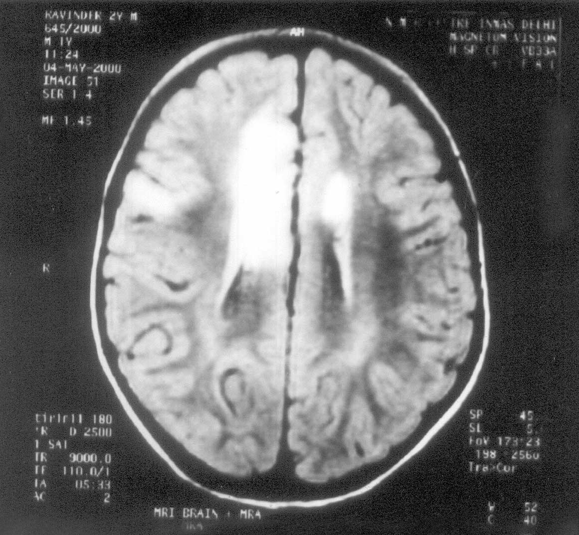
ADEM is a monophasic, polysymptomatic disorder involving central nervous system white matter. This immune mediated inflammatory process commonly follows viral infections, or may occur after vaccination. ADEM accounts for up to one third of all known cases of encephalitis. The polysymptomatic presentation reflects demyelinating lesions and consists of various combinations of motor, sensory, visual and cognitive symptoms. The clinicoradiological profile of 7 patients with acute disseminated encephalomyelitis is presented in this paper. Subjects and Methods The patients reported in this study were seen at Kalawati Saran Children’s Hospital, Delhi during the period of April 2000-March 2001. ADEM was suspected by development of acute neurological deficit occurring in close temporal relationship with a viral infection or vaccination and confirmed by typical radiological appearance on MRI. Cranial noncontrast and contrast enhanced CT was performed for all patients. MRI was performed for all patients with 1.5 T Magnetom vision of Siemens, Germany having a gradient strength of 25 MT /min. The non-contrast enhanced spin echo MRI was used to take axial, coronal, sagittal T1 weighted, T2 weighted spin echo images. In all planes slice thickness was 5 mm. Patients were followed up for a minimum of 6 months and follow up MRI was performed in 2 patients. Results The present study is a case series of seven patients diagnosed as ADEM by clinical presentation and radiological appearance and followed up for a minimum of 6 months. The cases have been summarized in Table I. The patients in our series, diagnosed as ADEM had very variable and polysymptomatic presentation. Three patients presented with focal presentation in the form of hemiparesis and focal seizures whereas there were cerebellar signs and generalized seizures in 1 patient. Three patients presented with altered sensorium. There was history of immunization in 2 patients (DPT/OPV in 1 and measles in other). CT Scan was normal or showed nonspecific hypo density. MRI clinched the diagnosis showing demyelination. This was best seen in T2 weighted sequence of MRI as areas of increased signal involving subcortical white matter, basal ganglia and deep grey matter (Fig. 1). Most of our patients had started showing improvement by the time the diagnosis was made, so steroids were administered in 3 patients only. TABLE 1 Clinical Profile of the Patients.
Discussion Acute disseminated encephalomyelitis is an acute inflammatory demyelinating disease of central nervous system. It is a monophasic polysymptomatic disease. It occurs due to immune mediated inflammatory process following infection or vaccination. ADEM usually follows viral infections. Viral pathogens commonly implicated are Measles, Varicella, Rubella, Mumps, Herpes Simplex, Herpes Zoster, Ebstein Barr Virus, Coxsackie Virus, Hepatitis B, HIV, and Human corona virus. Mycoplasma and Group A beta hemolytic streptococcus have also been incriminated. Symptoms usually follow 1-20 days of viral infection. ADEM may also occur following vaccinations with Measles, mumps, Rubella, Semple antirabies, influenza, small pox vaccine. Rarely it has also been reported following Organ transplantation, or as a paraneoplastic disorder in some cases of Leukemia and Non Hodgkin Lymphoma. The age group affected commonly is between 1-20 years. The hallmark of the disease is acute development of neurological signs accompanied by complaints of headache, fever, and alteration of sensorium. Since there is no specific presentation, it can mimic many other neurological disorders. The onset is abrupt with focal and multifocal signs reflecting cerebral (hemiparesis, aphasia) brainstem (Cranial nerve) and spinal cord (paraparesis) involvement. Focal manifestations in the form of hemiplegia, cerebellitis are more common in children than adults as observed in our cases, 4 out of 7 patients reported had focal presentation. The clinical features of post infectious and post vaccinal syndromes are quite similar except that additional peripheral nervous system involvement is more common in cases occurring after rabies vaccine. Radiculopathy has been reported in 82% of cases following nerve tissue rabies vaccine probably because the triggering vaccine contained peripheral nervous tissue(1). Cerebellar symptoms are very strongly associated with post varicella ADEM. In the presence of suggestive clinical features diagnoses is made by characteristic radiological appearance on MRI. MRI done early facilitates diagnosis and can lead to early treatment with the possibility of improved outcome(2). T2 weighted images reveal areas of increased signal intensity through out the CNS. T2 weighted images are more sensitive than T1 weighted images in detecting abnormalities. The lesions usually involve subcortical white matter and deep-seated grey matter including basal ganglia, thalamus and cerebellum(2). CT Scan may be normal or reveal areas of patchy low attenuation in the white matter. CT Scan lesions do not correspond to the extent or pattern of clinical disease(3). CT Scan was normal in 3 of our patients. CSF reports and EEG are non-specific. The most frequent findings are a mild mononuclear pleocytosis and mild elevation of proteins. Cerebrospinal fluid studies were normal in 5 patients in our series and showed lymphocytosis in 1 patient and raised protein in another patient. The pathological features of ADEM are perivenular inflammation and demyelination due to autoimmune mediated response to CNS myelin. Histologic staining for myelin basic protein and myelin oligodendrocyte glyco-protein demonstrates loss of these myelin proteins(4). There are very few studies of ADEM in pediatric age group in India. A study from south India by Murthy, et al. in all age groups found viral infections and Semple antirabies vaccine as the most common antecedent events. Multifocal form of presentation was seen in 56.5% of patients in that study(5). The other diseases that can present with similar clinical picture are viral encephalities, progressive multifocal lencoencephalopathy, mitochondrial encephalopathy, multiple sclerosis and even cerebrovascular accident. An early diagnosis by neuroimaging is important because treatment and prognosis depends on the exact diagnosis. ADEM and MS have many common clinical features including the possibility of a discrete infectious trigger. ADEM can sometimes be multiphasic and episodes may recur within 18 months of the initial attack without the patient going on to develop MS with long term follow up. These similarities make it more difficult to differentiate between these two diseases solely on clinical basis(6). ADEM is more commonly seen in younger age group whereas MS is more commonly seen in older age (25-30 years). MRI characteristically show asymmetrical periventricular lesions in MS whereas there are bilateral symmetrical sub cortical white matter and deep grey matter hypo densities in ADEM(7). Intrathecal IgG antibodies are seen in 70-90 percent in MS whereas in ADEM Intrathecal IgG are seen in less than 10 percent. Oligoclonal band is seen in 90-95% patients in MS which is persistent. In ADEM only transient Oligoclonal band is seen(8,9). ADEM is usually a self-remitting disease. Approximately 70% of the patients show complete recovery. Permanent neurological deficit remains in 10-20% of the patients. The mortality in the patients has been reported to be upto 30%. Up to 30% can have relapse if followed for longer period(10). Features that suggest a worse prognosis are coma and complicating seizures. Mild cognitive deficit remains in children who have otherwise recovered clinically and radiologically(11). Aside from supportive care many agents that target the immune status have been used and reported anecdotally to be effective. These include glucocorticords, adreno-corticotropin (ACTH), intravenous immunoglobulin, and plasmapheresis and glatiramer acetate. Glucocorticoids are considered effective because of their anti-inflammatory and immunomodulatory effect with an additional beneficial effect on cerebral edema. ACTH has also been reported by many studies, to be effective in ADEM(12) Intravenous immuno-globulin has been beneficial when used early in the disease course as well as when used late after failure to respond to gluco-corticoids(13,14). Conclusions ADEM is an acute inflammatory and demyelinating disease with a multifocal clinical presentation. Since there is no pathognomonic clinical or laboratory feature and CT scan is not very sensitive to diagnose this disorder; MRI should be considered early in patients with acute onset of unexplained encephalopathy or focal neurological deficit. It is a self-remitting disease with possible improved prognosis with immunomodulatory agents like steroids. Contributors: SM worked up the patients, reviewed the literature and drafted the manuscript. SA was the consultant in charge, provided the concept, co drafted the manuscript and revised it critically and is the guarantor of the study. RPT and AB performed the MRI and helped in interpretation and analysis of the radiological data and co drafted the manuscript. AS and VT contributed in data collection and co-drafting of the article. Funding: None. Competing Interests: None stated.
| ||||||||||||||||||||||||||||||||||||||||||||||||||||||||||||||||||||||||||||
|
References | ||||||||||||||||||||||||||||||||||||||||||||||||||||||||||||||||||||||||||||
|
. |
![]()