1.gif (1892 bytes)

Original Article

Indian Pediatrics 2005; 42:321-328

Long-term Outcome in Children with Primary Distal Renal Tubular Acidosis


Anurag Bajpai†, Arvind Bagga, Pankaj Hari, Aditya Bardia and Mukta Mantan

From the Division of Pediatric Nephrology, Department of Pediatrics, All India Institute of Medical Sciences, New Delhi 110 029, India.

Correspondence to: Dr. Arvind Bagga, Department of Pediatrics, All India Institute of Medical Sciences, Ansari Nagar, New Delhi 110 029, India.
E-mail: [email protected] 

Manuscript received: March 26, 2004, Initial review completed: July 2, 2004,
Revision accepted: November 16, 2004.

Abstract

Objective: To evaluate complications in adequately treated children with distal renal tubular acidosis (RTA) and to identify factors influencing their development. Design: Records of patients with primary distal RTA followed for 2 or more years at this hospital were reviewed. Case records were examined for age at onset of symptoms and at initiation of treatment, treatment details, follow-up and complications. Height, weight and growth velocity were expressed as standard deviation score (SDS) during different periods of follow-up. Regression analysis was performed to evaluate factors influencing increase in height and weight SDS. P value of less than 0.05 was considered significant. Results: Of 18 patients (eleven boys), the diagnosis was established at the median (range) age of 6 yr (1.5-13 yr). These patients were followed up for a median (range) period of 4 yr (2-18.5 yr). Short stature (height SDS <–2) was noted in all patients at the time of diagnosis with median (range) height SDS of –5.2 (–7.5 - –0.4). All patients had failure to thrive with median (range) weight SDS of –3 (–5.7 - –1.5). Height SDS increased by median (range) of 2 (1.2-5.5) to become –2.7 (–4.8 - –1.1) at last follow-up. Weight SDS increased by median (range) of 0.9 (–0.6 - 2.8) to become –2.4 (–4 - –0.5). Median (range) growth velocity SDS decreased from 3 (1-16) during first year of treatment to 1 (–0.3 - 7) at four years with an increase in mean height SDS by 1.3 during the first two years of treatment. Height SDS at last follow-up was not influenced by the age at initiation of treatment, follow-up duration, initial height SDS or severity of acidosis at diagnosis. Increase in height SDS correlated negatively with base excess and height SDS at diagnosis, and positively with follow-up duration on univariate analyses. Initial height SDS was the only factor that influenced increase in height SDS on multivariate analyses. Increase in weight SDS was negatively correlated with base excess and initial weight SDS with significant association with initial weight SDS, on multivariate analyses. All patients had rickets at the time of diagnosis with pathological fractures in four. Rickets resolved without treatment with vitamin D in seventeen patients after a median (range) period of 4 mo (1-12 mo). Eight patients (44.4%) had nephrocalcinosis at diagnosis. Twenty-four hour urine calcium excretion was higher in those with nephrocalcinosis (P = 0.01). Creatinine clearance remained normal in all except one who progressed to renal failure. Conclusions: There is a need for early diagnosis, appropriate treatment and regular follow-up of patients with distal RTA for improving outcome.

Keywords: Distal renal tubular acidosis, Growth, Nephrocalcinosis.

Distal renal tubular acidosis (RTA) is characterized by impaired renal tubular excretion of acid leading to hyperchloremic acidosis with inappropriately alkaline urine(1). The disease is associated with significant complications in the form of growth retardation, bone disease, nephro-calcinosis and development of chronic renal insufficiency(2). The effect of treatment on prevention of these complications is not clear. In particular there is paucity of published data on the effect of treatment on growth, a major cause of concern in these patients. Compromised final height despite adequate treatment has been reported(3-6). Identification of factors influencing the development of these complications is essential for improving the outcome of this disease. This retrospective study was performed to identify complications in adequately treated children with distal RTA and identify factors that predict their development.

Subjects and Methods

Records of patients with distal RTA followed for 2 or more years in the Renal Clinic of this hospital between the years 1984 to 2003 were reviewed. A patient was diagnosed to have distal RTA in the presence of all of the following (a) metabolic acidosis [total CO2 content (tCO2) <17.5 mEq/L], (b) normal blood anion gap (<14 mEq/L), (c) high urine pH (>5.5 with blood tCO2 <17.5 mEq/L), (d) positive urine net charge, (e) fractional excretion of bicarbonate <10% (after bicarbonate loading) and (e) urine to plasma carbon dioxide difference <10 mm Hg (after bicarbonate loading)(1). Children with impaired renal functions [creatinine clearance <50 mL/min/1.73m2, calculated using Schwartz formula(7)] and those where the disease was considered secondary to e.g., obstructive uropathy or systemic lupus were excluded.

Initial evaluation

Case records were evaluated for age at onset of symptoms and at initiation of treatment. Height or length was measured using stadiometer or infantometer. Height and weight was expressed as standard deviation score (SDS) with reference to NCHS standard(8). SDS was calculated as below:

           Subjects height-mean height
SDS = ————————————
           Standard deviation of mean

Blood levels of chloride, pH, bicarbonate, tCO2, urea, creatinine, calcium, phosphate and alkaline phosphatase were estimated. Calcium excretion was quantified on spot and 24-hour urine collections. Radiological evaluation included X-rays of wrists and long bones and ultrasound for kidneys. Rickets was defined as the presence of widened and irregular epiphyseal-metaphyseal junctions (6). Nephrocalcinosis was diagnosed in the presence of diffuse calcification of the renal pyramids(2).

Treatment and follow-up

Patients were treated with sodium-potassium citrate (Polycitra; 1 mL = 1 mEq sodium, 1 mEq potassium and 2 mEq bicarbonate). Dose of bicarbonate was adjusted to achieve plasma bicarbonate in the range of 20-24 mEq/L. Bicarbonate levels greater than 20 mEq/L was considered for adequacy of treatment. Dietary advice regarding salt restriction was given. Patients were followed up after every three months for the first 2 years and 6-12 months thereafter. Evaluation of anthropometeric parameters, clinical features and biochemical measurements were done at each visit. Patients with persistent hypercalciuria (>4 mg/kg/day) despite correction of acidosis were started on hydrochlorthiazide (2 mg/kg/day). X-ray wrist was done in patients with rickets every 3 months till healing was achieved. Patients with no nephrocalcinosis on first visit underwent annual ultrasound examination for kidneys.

Statistical analyses

Values are expressed as median (range) unless specified. SPSS version 10 was used for statistical analyses. Height and weight SDS at the initiation of treatment and at last follow-up were compared using paired t test. Linear regression analysis was done to evaluate factors influencing height and weight SDS at follow-up and increase in height and weight SDS. Variables examined included gender (boy or girl), age at initiation of treatment, severity of acidosis at the diagnosis (tCO2 and base excess), duration of treatment and initial SDS (height or weight). Variables found significant on univariate analyses were included in multivariate analyses. Features of subjects with and without nephrocalcinosis were compared using unpaired t test. P value of less than 0.05 was considered as significant.

Results

Of 18 patients (eleven boys), the onset of symptoms was at the age of 1.8 yr (3 mo-7.5 yr) and at diagnosis 6 yr (1.5-13 yr). All patients had polyuria (urine output > 6 mL/kg/hr) and hypokalemia at the time of diagnosis. Daily bicarbonate requirement decreased from 7.2 mEq/kg (4-8.2 mEq/kg) in the first 2 years of treatment to 3.4 mEq/kg (1.6-4.2 mEq/kg) thereafter. Patients were followed up for 4 yr (2-18.5 yr). Correction and normalization of levels of bicarbonate, tCO2 and base excess was achieved in 16 patients. One of the 2 patients with persistent metabolic acidosis had poor compliance; the other showed renal insufficiency.

Growth parameters

Short stature (height SDS <–2) was noted in all patients at the time of diagnosis with height SDS of –5.2 ((7.5-0.4). Nine patients (50%) had height SDS less than –5. No significant correlation of height SDS at initiation of treatment with age at diagnosis (r = –0.33, P = 0.2) or level of tCO2 (0.34, P = 0.17) was observed. All patients had failure to thrive with weight SDS of –3 (–5.7 - –1.5). The median weight for height was 94% (78-120%), it was greater than 80% in all but one subject.

The median height SDS increased by 2 (–1.2 - 5.5) to become –2.7 (–4.8 - 1.1) at last follow-up (Table I). Height SDS increased in all patients with an exception of two patients with persistent metabolic acidosis (Table II). Height SDS at last follow-up was greater than –5 in all and greater than –2.5 in eight patients (44.4%). The median weight SDS increased by 0.9 (–0.6 - 2.8) to become –2.4 (–4 - –0.5) at last follow-up.

TABLE I
Follow-up Details of Individual Patients.
No. Age */ Sex Follow-up HSDS
initial
HSDS
end
HSDS
increase
Nephrocalcinosis
Bone
disease
1
8 yr M
18.5 yr
–6.8
–2.2
4.6
Yes
R
2
3.5 yr M
3 yr
–2.7
–2.5
0.2
Yes
R
3
10 yr M†
4.6 yr
–3.8
–4.8
–1
Yes
R
4
6 yr M
18.6 yr
–7.5
–3.3
4.2
No 
R, Fr
5
7.5 yr M 
3 yr
–4.8
–2.7
2.1
No
R
6
2 yr F
7.5 yr
–6.8
–1.3
5.5
No
R
7
6 yr M
4 yr
–5.5
–4
1.5
No
R,Fr
8
10 yr M
4 yr
–5.8
–3.6
2.2
No
R
9
1.5 yr F
4 yr
–3.4
–3.2
0.2
No
R
10
3 yr F
3.6 yr 
–3.9
–2
1.9
No
R
11
13 yr M
3 yr
–5.7
–3.9
1.8
No
R
12
2.2 yr F‡
4 yr
–0.4
–1.6
-1.2
No
R
13
2.5 yr M
2 yr
–5.8
–4.8
1
Yes
R
14
6 yr M
5.5 yr
–3.5
–1.6
1.9
Yes
R, Fr
15
6 yr F
4 yr
–2.9
1.1
4
Yes
R, Fr
16
11 yr F
3 yr
–5.4
–2.2
3.2
Yes 
R
17
7 yr F
3
–5.5
–2.7
2.8
Yes
R
18
6 yr M
4.2
–5
–3.5
1.5
No
R

* At diagnosis; † Developed renal failure 2 years after diagnosis; ‡ No improvement in metabolic acidosis despite adequate treatment; M-male, F-female, R-rickets, Fr-fracture, HSDS-Height standard deviation score.

TABLE II

Height and Weight SDS at Initiation of Treatment and at
 Last Follow-up.
Parameter
Initial 
End
P
Height SDS
–5.2 (–7.5 - –0.4) 
–2.7 (–4.8 - 1.1) 
<0.0001
Weight SDS
–3 (–5.7 - –1.5)
–2.4  (–4 - –0.5)
<0.0001
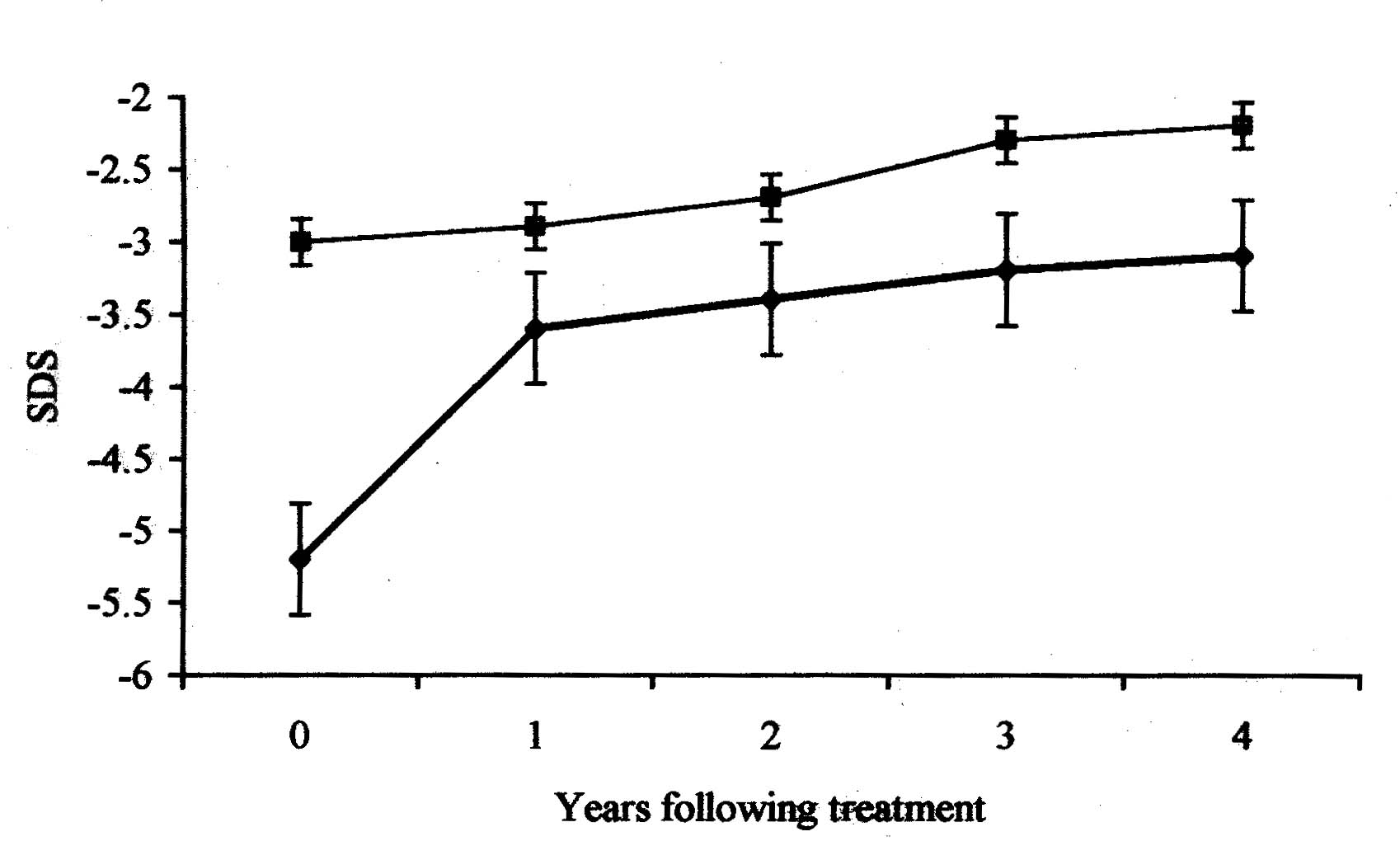
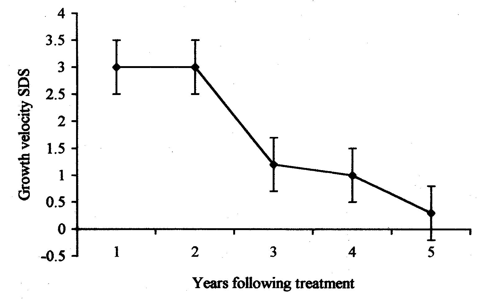
Expressed as median (range), SDS = Standard deviation score.

The pattern of height, weight and growth velocity with respect to duration of treatment is shown in Figs. 1 and 2. Growth velocity decreased from 10 cm/yr (4-16 cm/yr) during the first year of treatment to 5 cm/yr (4-6 cm/yr) at four years. This was associated with an increase in height SDS by 1.3 during the first two years of treatment. Height SDS increased by 0.7 during subsequent follow-up.

Fig. 1. Line diagram showing trend of median height (lower line) and weight SDS (upper line) during treatment.

Fig. 2. Line diagram showing trend of median growth velocity SDS during treatment.

Factors influencing growth are summarized in Tables III and IV. Height SDS at the end of treatment was not influenced by age at initiation of treatment, follow-up duration, initial height SDS or severity of acidosis at diagnosis. Increase in height SDS correlated negatively with base excess and height SDS on diagnosis and positively with follow-up duration on univariate analyses. On multivariate analysis initial height SDS was the only factor that influenced increase in height SDS. Increase in weight SDS was negatively correlated with base excess and initial weight SDS, with significant association with initial weight SDS on multivariate analyses. A trend of association of age at diagnosis and weight SDS at last follow up was observed (P = 0.07).

TABLE III

Factors Influencing Height and Weight SDS on Univariate Analyses.
		
Variable Height SDS Weight SDS
Last follow-up Increase Last follow-up Increase
r P r P r P r P
Age diagnosis yr 
–0.33
0.18
0.07
0.77
–0.54
0.02
–0.12
0.65
Base excess mEq/L*
–0.17
0.51
–0.48
0.04
0.29
0.24
–0.58
0.01
Bicarbonate mEq/L*
–0.08
0.75 
–0.38
0.12
0.11
0.66
–0.39
0. 11
Follow-up yr
0.02
0.94
0.52
0.03
0.65
0.003
0.31
0.20
Initial SDS
0.33
0.19
–0.73
0.001
–0.06
–0.80
–0.64
0.004
* At the time of diagnosis, r - correlation coefficient, SDS - standard deviation score.
TABLE IV

Multivariate Analyses for Factors Influencing Height and Weight SDS.
  Increase height SDS Increase weight SDS
Variable Coefficient*  P Coefficient* P
Base excess
–0.21
0.32
–0.27
0.32
Initial SDS
–0.59
0.01
–0.46
0.10
Follow-up yr
0.10
0.64
Adjusted R2†
49.4%
44.9%

* Regression coefficient provides measure of influence of the independent variable on the dependent variable. For example increasing follow-up duration by one year is expected to increase height SDS by 0.1.
† Adjusted R2 provides a measure of variation explained by the model used for regression analyses. For example base excess, initial height SDS and follow-up period explain 49.4% of the variation observed in increase in height SDS.

Bone disease

All patients had radiological features of rickets at the time of diagnosis with pathological fractures in four. Serum alkaline phosphatase was normal in 10 patients (62.5%). Radiological findings of rickets resolved in seventeen patients after 4 (1-12) months, without treatment with vitamin D.

Nephrocalcinosis

Eight patients (44.4%) had radiological evidence of nephrocalcinosis. Twenty-four hour urine calcium excretion was higher in those with nephrocalcinosis than others (P = 0.01); no difference with regards to the age at diagnosis or severity of acidosis was found (Table V). No patient showed renal calcification during follow-up. Five of the 10 patients with hypercalciuria at diagnosis showed reduction in calcium excretion following alkali treatment. Treatment with hydrochlorthiazide was used in the remaining subjects.

TABLE V
Comparison of Patients with and Without Nephrocalcinosis.
  Nephrocalcinosis P
Factor Yes No
 Age at diagnosis (yr) 
6.5 (0.5-7) 
6.0 (0.3 - 7.5)
0.53
 24 hour urine calcium (mg/kg/day)
7.6 (6-20) 
3.0 (2.5 - 5)
0.01
 Serum bicarbonate (mEq/L)
14.5 (9.5-18) 
12.3 (10.2 -16)
0.40
 Base excess (mEq/L)
–12.8 (–17- –6) 
–15.0 (–19.7 - –8) 
0.96
 tCO2 (mEq/L)
12.0 (10 -18.2) 
12.5 (10.6 - 16.4)
0.32
 Expressed as median (range).

Renal functions

Creatinine clearance remained normal in all except one patient. This boy diagnosed at the age of 10 years showed bilateral medullary nephrocalcinosis with normal renal functions at diagnosis. Despite adequate treatment the creatinine clearance declined to 20 mL/min/1.73 m2 2 years later.

Discussion

The present study shows that appropriate treatment in distal RTA is associated with significant increase in height SDS; final height SDS however remains compromised. There is a paucity of similar published data on growth in children with distal RTA. Rodriguez, et al. (5 patients) and Santos, et al. (24 patients) demonstrated compromised height in children with distal RTA(4, 5). More recently Caldas, et al., showed compromised stature in 28 patients with distal RTA. The mean height SDS was –1.1 for patients diagnosed during infancy and –2 for those diagnosed later(6). The extent of height compromise (final mean height SDS of –2.6) in the present study is greater compared to previous studies. This may be related to delayed diagnosis, greater incidence of bony deformities and lower genetic potential of these patients.

An understanding of factors influencing height outcome in subjects with RTA is necessary. In this study, initial height SDS was the only factor that negatively correlated with increase in height SDS. The duration of treatment and severity of acidosis at the time of diagnosis were also found to influence growth on univariate but not on multivariate analysis. This suggests that maximum gain in height SDS was observed in patients who had greatest height compromise at the initiation of treatment. Our findings imply that there is a trend of seeking target height after correction of acidosis. Our failure to demonstrate the effect of age at diagnosis on final height may be related to delayed diagnosis in all the subjects. Majority of our patients were diagnosed after two years of age. It appears that this is a crucial window period for intervention beyond which height is already compromised. These findings are similar to those of Caldas, et al.(6) who observed compromised final height in patients diagnosed after infancy and emphasize the need of early diagnosis and treatment of this condition. Nutritional status influences growth; normal weight for height in majority of our subjects indicates that RTA and not malnutrition was the major factor responsible for growth retardation in our subjects.

The pattern of height and growth velocity SDS in our study shows that maximum catch up growth occurs during the first two years of treatment with a decline in growth velocity and increase in height SDS thereafter. Similar findings were demonstrated by others who found that catch up growth was limited to the first two years of treatment(6). This coupled with the fact that increase in height SDS during the first two years of treatment significantly correlated with overall increase in height SDS emphasizes the need for improved management during this crucial period.

A high incidence of bony deformities and pathological fractures in this study is distinct from previous reports where rickets was uncommon in children with distal RTA(9). Demonstration of normal levels of vitamin D metabolites and resolution of rickets without treatment with vitamin D suggest a minor role for its altered metabolism in the pathogenesis of bone disease in distal RTA(10,11). It has been suggested that vitamin D status of patients plays a role in determining the extent of bony deformities(6). It is possible that low vitamin D stores in our patients explain the presence of rickets and pathological fractures. A significant proportion of patients with rickets in this study had normal levels of serum alkaline phosphatase. Studies have shown that bone formation is suppressed in patients with distal RTA. Levels of serum alkaline phosphatase, a marker of bone formation, are therefore expected to be normal in these subjects.

The incidence of nephrocalcinosis in our study is similar to previous studies(2,6). Prevention of renal calcification is essential for preservation of renal functions in these patients. Hypercalciuria was the only factor that predicted the development of nephro-calcinosis, with no influence of age at diagnosis or severity of acidosis. Our findings emphasize the need for close monitoring and appropriate management of hypercalciuria. While other studies have shown that nephro-calcinosis may develop during treatment(6), we did not find the recurrence in our subjects. Adequate treatment with alkali supplements combined if required, with thiazide diuretics, may thus prevent the development and worsening of nephrocalcinosis.

The findings of our study reiterate the need for early diagnosis, appropriate treatment and regular follow-up of patients with distal RTA for improving outcome. Significant height compromise despite adequate treatment emphasizes the need for studies directed at evaluating factors other than metabolic acidosis that are responsible for growth in these patients.

Contributiors: MM, PH and AB were involved in management of patients. AnB and AdB planned the study and collected data. AnB performed the literature review and drafted the manuscript. AB designed the study and critically reviewed the manuscript and will act as its guarantor.

Competing interest: None.

Funding: None.

Key Messages


• Treatment of children with distal RTA is associated with significant improvement in growth parameters, final height however remains compromised in these children.

• First two years of therapy are crucial and associated with greatest improvement in growth parameters.

• Early diagnosis and management of hypercalciuria is helpful in preventing the progression of renal calcification and development of chronic renal insufficiency.


References


1. Herrin TJ. Renal tubular acidosis. In: Pediatric Nephrology, 4th edn. Eds. Baratt TM, Avner ED, Harmon WE. Baltimore, Williams & Wilkins, 1999, pp 565-583.

2. Caruana RJ, Buckalew VM Jr. The syndrome of distal (type 1) renal tubular acidosis. Clinical and laboratory findings in 58 cases. Medicine 1988; 67: 84-99.

3. Nash MA, Torrado AD, Griefler I, Spitzer A, Edelman CM Jr. Renal tubular acidosis in infants and children. Clinical course, response to treatment and prognosis. J Pediatr 1972; 80: 738-748.

4. Rodriguez-Soriano J, Vallo A, Castillo G, Oliveros R. Natural history of distal renal tubular acidosis treated since infancy. J Pediatr 1982; 101: 669-676.

5. Santos F, Chan JC. Renal tubular acidosis in children. Diagnosis, treatment and prognosis. Am J Nephrol 1986; 6: 289-295.

6. Caldas A, Broyer M, Dechaux M, Klienknecht C. Primary distal tubular acidosis in childhood: Clinical study and long term follow up of 28 patients. J Pediatr 1992; 121: 233-241.

7. Schwartz GJ, Haycock GB, Edelman CM, Spitzer A. A simple measure of glomerular filtration rate in children derived from body length and plasma creatinine. Pediatrics 1976; 58: 259-263.

8. Hamill PV, Dizd TA, Johnson CL. NCHS curves for children, birth-18 yr. United States Vital Health Statistics 1977; I-IV: 1-74.

9. Brenner RJ, Spring DB, Sebastian A, McSherry EM, Genant HK, Palubinskas AJ, et al. Incidence of radiographically evident bone disease, nephrocalcinosis and nephrolithiasis in various types of renal tubular acidosis. N Engl J Med 1982; 307: 217-221.

10. Cunningham J, Fraher LJ, Revell PA, Papappoulos SE. Chronic acidosis with metabolic bone disease - Effect of alkali on bone morphology and vitamin D metabolism. Am J Med 1982; 73: 199-204.

11. Chesney RW, Kaplan BS, Phelps M, DeLuca HF. Renal tubular acidosis does not alter circulating values of calcitriol. J Pediatr 1984; 104: 51-55.


Home

Past Issue

About IP

About IAP

Feedback

Links

 Author Info.

  Subscription